Session Information
Session Type: Poster Session A
Session Time: 10:30AM-12:30PM
Background/Purpose: Rheumatoid arthritis (RA) is a female predominant disease characterized by chronic inflammatory erosive joint disease. Androgen, the dominant sex hormone in males, is anti-inflammatory and protective against bone loss (1,2). We have previously shown that androgen limits erosions in a TNF-mediated murine model of RA. Here, we investigated the cellular and molecular targets of androgen and hypothesized that androgen negatively regulates the process of osteoclastogenesis in precursor cells.
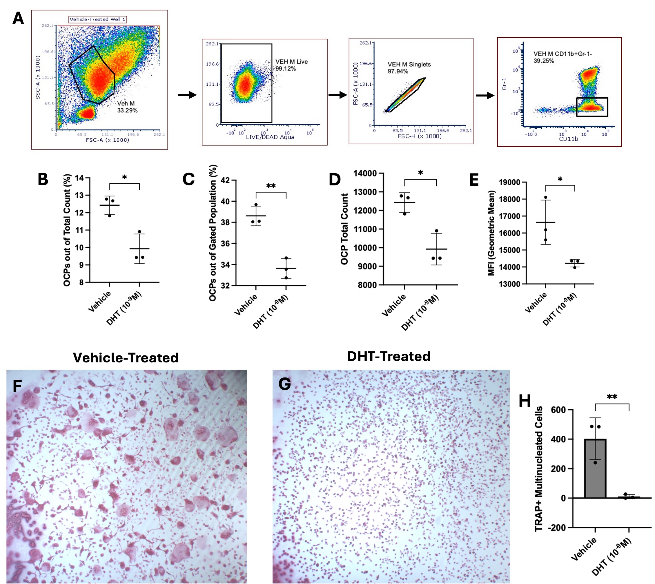
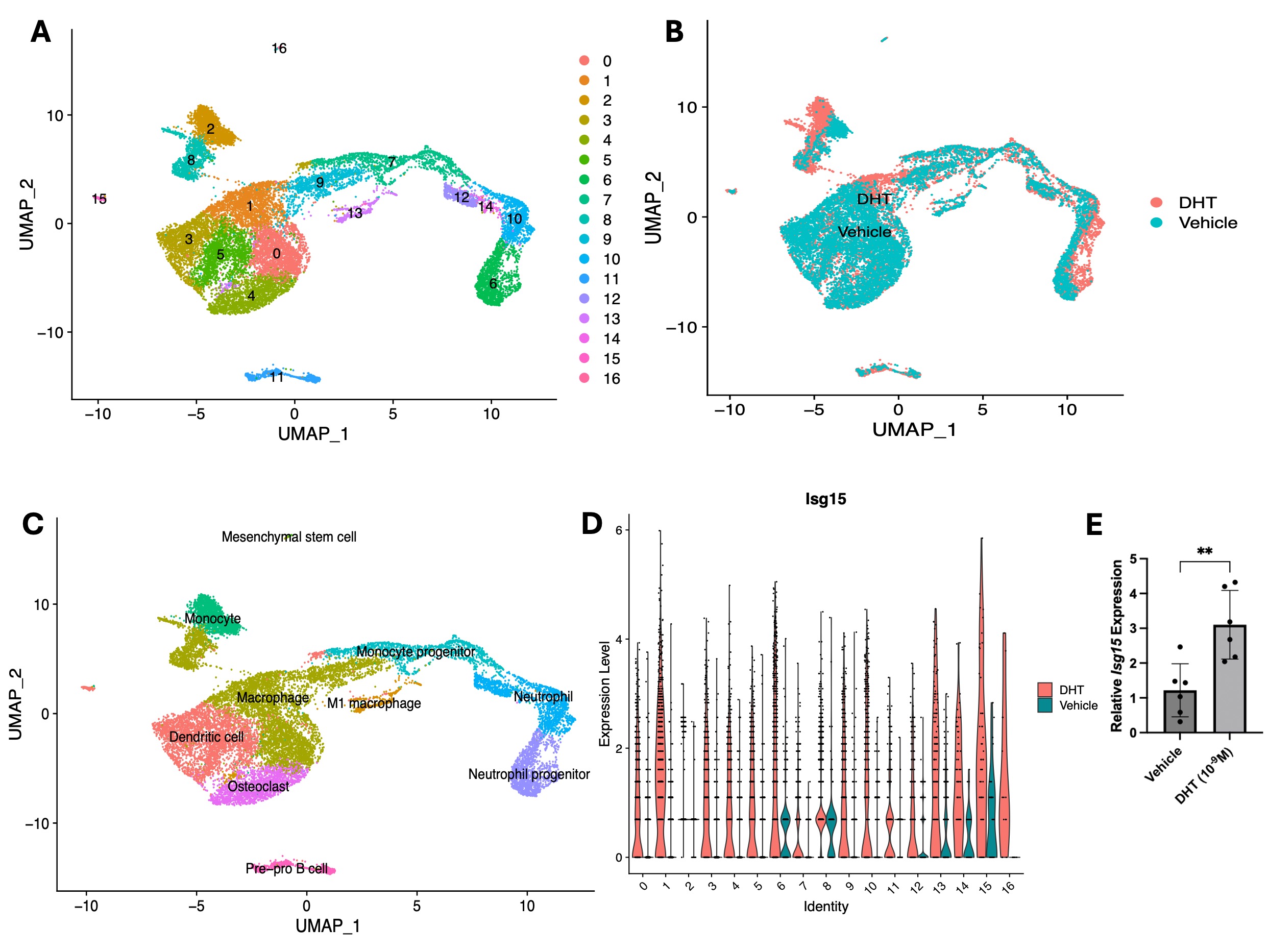
Methods: Bone marrow derived (BMD) cells were harvested from a 3-month-old male C57BL/6J mouse and plated in media containing either 10-9M of 5ɑ-dihydrotestosterone (DHT) or vehicle control, M-CSF, and human TNF-alpha (hTNFɑ) for 48 hours. Adherent cells were collected for flow cytometry analysis and fluorescence-activated cell sorting (FACS). Osteoclast precursors (OCPs) were defined as myeloid-like CD11b+Gr-1- cells and were replated in media with RANKL, M-CSF, and hTNFɑ. After osteoclast formation, cells were fixed and TRAP-stained. The total number of osteoclasts (≥3 nuclei, TRAP+) was quantified in each well (n = 3 wells/condition). We then performed single-cell RNA sequencing (scRNA-seq) on our culture system. BMD cells from a 3-month-old male mouse were plated with 10-9 DHT or vehicle and M-CSF. After 48 hours, RANK-L, M-CSF, and hTNFɑ were added to media and cells were incubated for 24 hours. Adherent cells were captured with the 10X Genomics Chromium system. Cell typing was determined by the single cell mouse cell atlas and computational analysis was performed in the Seurat R package. Using the same cell culture protocol, RT-qPCR was performed on DHT and vehicle-treated samples to compare gene expression. Values are reported as mean ± standard deviation and compared with unpaired t-tests.
Results: DHT-treated cells had a significantly decreased OCP population compared to vehicle-treated cells (DHT = 9.93 ± 0.85%, Vehicle = 12.43 ± 0.53%) (Fig. 1B-E). DHT-treated cells also had significantly less osteoclasts in culture (DHT = 11 ± 14 cells, Vehicle = 403 ± 142 cells) (Fig. 1F-H). In scRNA-seq, 17 clusters were defined in the DHT and vehicle-treated samples that included myeloid lineage cells and other cell types in osteoclast culture (Fig 2A-C). The top differentially expressed gene (DEG) in the DHT-treated sample in majority of clusters was interferon stimulated gene 15 (Isg15) (Fig 2D). Relative Isg15 expression was also significantly increased in DHT-treated OCPs (Fig 2E).
Conclusion: Androgen-treated cells had significantly smaller OCP populations and less mature osteoclasts compared to vehicle-treated cells. This suggests that androgen targets OCP populations and inhibits osteoclastogenesis. Isg15 has been suggested to negatively regulate osteoclastogensis in precursor cells (3). Increased Isg15 expression in DHT-treated samples suggests that androgen may regulate osteoclast differentiation through interferon signaling. Further studies will include examining how androgen specifically targets Isg15 expression and the role of Isg15 in osteoclastogenesis.
1) Traish A et al. J Clin Med 7(12): 549. 2018.
2) Chen JF et al. Cells 8(11) 2019.
3) MacLauchlan et al. Proc Natl Acad Sci U S A 120(15) 2023.
To cite this abstract in AMA style:
Chen K, Benoodt L, Christof M, Rahimi H. Elucidating Androgen Effects on Osteoclast Precursor Populations and Osteoclastogenesis [abstract]. Arthritis Rheumatol. 2024; 76 (suppl 9). https://acrabstracts.org/abstract/elucidating-androgen-effects-on-osteoclast-precursor-populations-and-osteoclastogenesis/. Accessed .« Back to ACR Convergence 2024
ACR Meeting Abstracts - https://acrabstracts.org/abstract/elucidating-androgen-effects-on-osteoclast-precursor-populations-and-osteoclastogenesis/


