
Background/Purpose : CNS involvement in Behçet’s disease, usually called neuro-Behçet’s disease (NB), can be classified into acute NB (ANB) and chronic progressive NB (CPNB) based upon the differences in clinical courses and responses to corticosteroid treatment. Previous studies demonstrated that brainstem atrophy was significantly more frequently observed in CPNB than in ANB or non-NB. Since the presence of brainstem atrophy depends on the anecdotal judgment by expert radiologists, more objective definition of brainstem atrophy is required. The present study was designed to examine whether quantitative analysis of the brainstem areas on magnetic resonance imaging (MRI) scans might be useful for early diagnosis as well as for evaluation of the disease activity of CPNB.
Methods : MRI scans recorded at the diagnosis and at various periods thereafter were evaluated in patients with ANB (n=10), CPNB (n=10), non-NB (n=8), and NPSLE (n=8). MRI scans of age- and sex- matched control patients for CPNB (non-inflammatory CNS diseases [NID]) (n=10) were also studied. The areas of midbrain tegmentum and pons were measured on mid-sagittal sections of T1-weightened images of brain MRI using image analysis software Image J (ver.1.45, National Institutes of Health: NIH, U.S. [http://imagej.nih.gov/ij/download.html]).
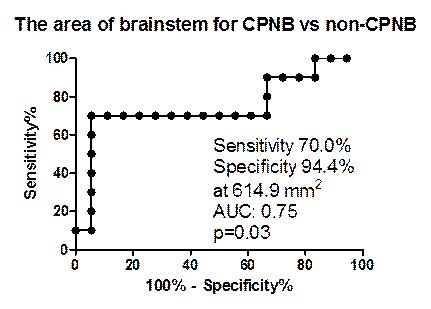
Results : The areas of midbrain tegmentum (ANB: 145.9 ± 20.1 mm2 [mean ± SD], CPNB: 125.6 ± 18.0 mm2, non-NB: 133.0 ± 16.4 mm2, NPSLE: 133.4 ± 19.7 mm2, NID: 151.1 ± 11.8 mm2) as well as those of pons (ANB: 540.1 ± 87.2 mm2, CPNB: 482.0 ± 91.3 mm2, non-NB: 540.3 ± 40.2 mm2, NPSLE: 517.3 ± 47.1 mm2, NID: 574.1 ± 43.4 mm2) were found to be lower in CPNB than those in the other 4 groups. On receiver operating characteristic (ROC) analysis, the sensitivity / specificity of the areas of midbrain tegmentum, pons and brainstem (midbrain tegmentum + pons) for diagnosis of CPNB against non-CPNB (ANB + non-NB) were 80.6% / 66.7% at cut-off value of 134.0 mm2, 70.0% / 77.8% at cut-off value of 483.7 mm2 and 70.0% / 94.4% at cut-off value of 614.9 mm2 respectively (figure). The time kinetics analysis demonstrated that brainstem atrophy progressed most markedly during the first 2 years from the initial diagnosis of CPNB. Finally, brainstem atrophy progressed at significantly greater degree in CPNB patients with continuous elevation of cerebrospinal fluid (CSF) IL-6 (>20pg/ml) compared with those in whom CSF IL-6 levels were kept bellow 20 pg/ml.
Conclusion : These results confirm that brainstem atrophy is one of the characteristic features of CPNB. Moreover, the data suggest that quantitative analysis of the brainstem areas on MRI scans might be effective for early diagnosis as well as for evaluation of the disease activity of CPNB. Finally, it is also suggested that brainstem atrophy might progress due to CNS inflammation mediated by IL-6 during the early phase of CPNB.
Disclosure:
H. Kikuchi,
None;
M. Takayama,
None;
Y. Kimura,
None;
K. Asako,
None;
H. Kono,
None;
Y. Ono,
None;
S. Hirohata,
None.
« Back to 2012 ACR/ARHP Annual Meeting
ACR Meeting Abstracts - https://acrabstracts.org/abstract/efficacy-of-quantitative-analysis-of-brainstem-atrophy-on-magnetic-resonance-imaging-for-diagnosis-of-chronic-progressive-neuro-behcets-disease/