Session Information
Session Type: Poster Session A
Session Time: 8:30AM-10:30AM
Background/Purpose: The disease activity assessment in c-TA is a challenge in clinical practice, especially in patients who are under immunosuppression. Our aim was to perform comparison between positron emission tomography (PET) and magnetic resonance angiography (MRA) findings in childhood-onset Takayasu’s arteritis (c-TA) patients.
Methods: A three-center cross-sectional study with 17 children and adolescents who fulfilled the EULAR/PRINTO/PReS criteria for c-TA was conducted. The patients underwent 18F-FDG-PET/MRI imaging (SIGMATM PET/MR, GE Healthcare, Milwakee-MI, USA). Positive MRA consisted of arterial wall thickening with contrast-enhancement, whereas positive PET was defined as arterial wall uptake higher than the normal liver degree of FDG uptake. Patients were divided in three groups according to the different combinations of imaging findings as following: group 1 (PET+MRA+), group 2 (PET+MRA-) and group 3 (PET-MRA+).
Results: Seventeen c-TA patients (65% female) were included. Mean disease duration was nine ±3.6 years. PET identified high-grade 18F-FDG arterial wall uptake in 15 (88.2%) patients and MRA detected thickening with contrast-enhancement in 10 (58.8%). Nine patients were classified as group 1, six as group 2 and 1 as group 3. One patient had no vessel-wall changes in both images. Median of metabolic inflammatory volume (MIV) value was significantly higher in group 1 compared to group 2 (2346 vs. 1177 cm3; p=0.036) (Table 1). Fifty-four (19%) from 284 available arterial segments presented vessel-wall changes in one or both imaging. Positive PET was found in 35 (12.3%) and positive MRA in 26 (9.2%) vessels (Table 2). Positive findings in vessel-wall were concordant between PET and MRA exams in seven (2.5%) and were not concordant in 47 (16.5%) arterial segments.
Conclusion: Our study demonstrated that 18F-FDG-PET/MRI is a new exam that adds metabolic information to anatomic image in c-TA patients, and may help therapeutic decision, especially for those during immunosuppressive withdrawal.
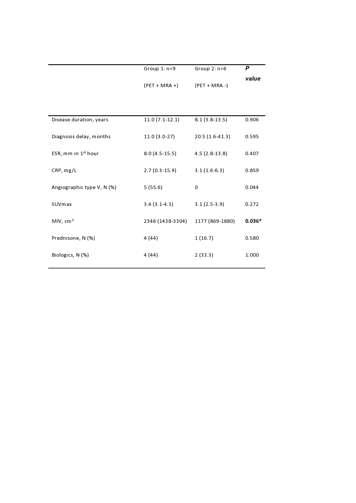
c-TA: childhood-onset Takayasu’s arteritis; ESR: erythrocyte sedimentation rate; CRP: C reactive protein; PET: positron emission tomography; MRA: magnetic resonance angiography; SUVmax: maximum standardized uptake value; MIV: metabolic inflammatory volume. Numerical values are presented in median (IQR). *1 patient had no vessel wall changes in MRA or PET imaging and 1 patient had vessel-wall changes in MRA but no vessel-wall changes in PET.

c-TA: childhood-onset Takayasu’s arteritis; PET: positron emission tomography; MRA: magnetic resonance angiography; VS: visual score; T+E: thickening + enhancement; RCC: right common carotid; LCC: left common carotid * Vessel-wall changes in PET and MRA that have undergone surgical intervention (5 arterial segments) were not considered.
To cite this abstract in AMA style:
Clemente G, Souza A, Leão Filho H, Coelho F, Buchpiguel C, Lima M, Pioto D, Fraga M, Sakamoto A, Carneiro C, Pereira R, Aikawa N, Silva C, Campos L, Astley C, Gualano B, Terreri M. Does 18F-FDG-PET/MRI Add Metabolic Information to Anatomic Image in Childhood-onset Takayasu’s Arteritis Patients? A Multicenter Case Series [abstract]. Arthritis Rheumatol. 2021; 73 (suppl 9). https://acrabstracts.org/abstract/does-18f-fdg-pet-mri-add-metabolic-information-to-anatomic-image-in-childhood-onset-takayasus-arteritis-patients-a-multicenter-case-series/. Accessed .« Back to ACR Convergence 2021
ACR Meeting Abstracts - https://acrabstracts.org/abstract/does-18f-fdg-pet-mri-add-metabolic-information-to-anatomic-image-in-childhood-onset-takayasus-arteritis-patients-a-multicenter-case-series/