Session Information
Date: Monday, November 14, 2022
Title: Abstracts: Spondyloarthritis Including PsA – Diagnosis, Manifestations, and Outcomes II: Imaging
Session Type: Abstract Session
Session Time: 4:30PM-6:00PM
Background/Purpose: Presence of vertebral corner inflammation (VCI) increases the likelihood of a new syndesmophyte in the same vertebral corner (VC) in patients with r-axSpA. It was suggested that subsequent vertebral corner fat deposition (VCFD) partially explains this effect. However, this has not been formally tested. We assessed how much of the effect of VCI on the development of new syndesmophytes is explained by new VCFD.
Methods: Two datasets (SIAS cohort, ASSERT clinical trial) were analyzed. Patients with r-axSpA were assessed at baseline (T0), an intermediate visit (T1) (SIAS: 1 year; ASSERT: 24 weeks) and the end of follow-up (T2) (SIAS: 2 years; ASSERT: 102 weeks). Syndesmophytes were assessed on whole spine low dose CT (SIAS) or spinal radiographs (ASSERT) at T0 and T2 and considered present if seen by 2 of 2 readers. VCI (T0) and VCFD (T0 and T1) on spinal MRI were present if seen by ≥2 of 3 readers (SIAS) or 2 of 2 readers (ASSERT). VCs with VCFD or a syndesmophyte at baseline were excluded. We used the counterfactual approach (Pearl, 2011) to decompose the total effect of VCI at T0 (binary exposure) on the formation of a new syndesmophyte in the same VC at T2 (binary outcome) into the effect that is explained (natural indirect effect, NIE) and the effect that is not explained (natural direct effect, NDE) by new VCFD (binary mediator) at T1. Because there was no interaction between the exposure and mediator (p=0.88 for SIAS; p=0.82 for ASSERT), the average NIE (aNIE) and average NDE (aNDE) are reported. The aNIE, aNDE and total effect, expressed as absolute increase in risk, were estimated in R using the ‘mediation’ package, which takes into account the 2-level structure of the data (VCs nested within patients).
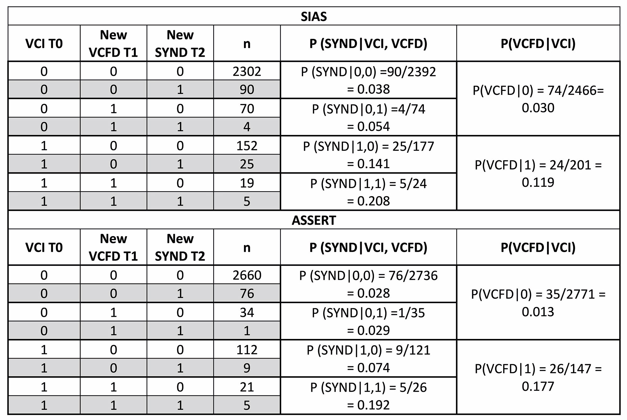
Results: In total, 49 patients (2,667 corners) in SIAS and 168 patients (2,918 corners) in ASSERT were included. A new syndesmophyte occurred at T2 in 124/2,667 (5%) corners in SIAS and 91/2,918 (3%) corners in ASSERT (Table). New VCFD at T1 was also uncommon (SIAS: 4%; ASSERT: 2%), but occurred more often in corners with (SIAS: 12%; ASSERT: 18%) than without VCI at T0 (SIAS: 3%; ASSERT: 1%). Applying the mediation formula, in SIAS, the presence of VCI at T0 increased the probability of a new syndesmophyte in the same VC at T2 by 9.3% [total effect (95% CI)=9.3% (4.5; 15.0)]. There was only a 0.2% increase in this probability that was mediated by the formation of new VCFDs at T1 [aNIE=0.2% (-0.4; 1.0)]. In contrast, 9.1% of the increase in probability remained unexplained [aNDE=9.1 (4.3; 15.0)]. This means that only 2% (0.2/9.3) of the total effect of VCI on the formation of new syndesmophytes was explained by new VCFD [% mediated=2.0% (-4.1; 13)]. In ASSERT, the total effect was somewhat lower than in SIAS [total effect=7.3% (2.0; 16.0)], and again the aNIE was small [aNIE=0.8% (-0.2; 3.0)], and the aNDE composed most of the total effect [aNDE=6.5% (1.3; 14.0)]. The proportion of the total effect explained by VCFD (0.8/7.3=10% (-3.1;44)) was larger than in SIAS but still non-significant.
Conclusion: In these two datasets we see that VCI only infrequently leads to syndesmophyte formation via visible VCFD.
VCI, vertebral corner inflammation; VCFD, vertebral corner fat deposition
To cite this abstract in AMA style:
Stal R, Sepriano A, Ramiro S, van Gaalen f, Machado P, Baraliakos X, de Hooge M, van den Berg R, Reijnierse M, Braun J, Landewé R, van der Heijde D. Do Fatty Lesions Explain the Association Between Inflammation and New Syndesmophytes in Patients with Radiographic Axial Spondyloarthritis? [abstract]. Arthritis Rheumatol. 2022; 74 (suppl 9). https://acrabstracts.org/abstract/do-fatty-lesions-explain-the-association-between-inflammation-and-new-syndesmophytes-in-patients-with-radiographic-axial-spondyloarthritis/. Accessed .« Back to ACR Convergence 2022
ACR Meeting Abstracts - https://acrabstracts.org/abstract/do-fatty-lesions-explain-the-association-between-inflammation-and-new-syndesmophytes-in-patients-with-radiographic-axial-spondyloarthritis/


