ACR Meeting Abstracts

ACR Meeting Abstracts

  • Meetings
    • ACR Convergence 2024
    • ACR Convergence 2023
    • 2023 ACR/ARP PRSYM
    • ACR Convergence 2022
    • ACR Convergence 2021
    • ACR Convergence 2020
    • 2020 ACR/ARP PRSYM
    • 2019 ACR/ARP Annual Meeting
    • 2018-2009 Meetings
    • Download Abstracts
  • Keyword Index
  • Advanced Search
  • Your Favorites
    • Favorites
    • Login
    • View and print all favorites
    • Clear all your favorites
  • ACR Meetings

Abstract Number: 1230

Differences in Nailfold Capillary Morphology Distinguish Juvenile Dermatomyositis Patients That Are Myositis-Specific Autoantibody Positive

Nicholas McClellan, Sarah Vandenbergen, Sophia Matossian, J. Michelle Kahlenberg and Jessica Turnier, University of Michigan, Ann Arbor, MI

Meeting: ACR Convergence 2023

Keywords: dermatomyositis, Pediatric rheumatology, Systemic lupus erythematosus (SLE)

  • Tweet
  • Click to email a link to a friend (Opens in new window) Email
  • Click to print (Opens in new window) Print
Session Information

Date: Monday, November 13, 2023

Title: (1221–1255) Pediatric Rheumatology – Clinical Poster II: Connective Tissue Disease

Session Type: Poster Session B

Session Time: 9:00AM-11:00AM

Background/Purpose: Juvenile dermatomyositis (JDM) is characterized by multiorgan vasculopathy, and degree of vasculopathy can indicate more severe disease. Nailfold capillaroscopy is a non-invasive method to directly evaluate abnormalities in the microvasculature. While the majority of children with JDM present with nailfold capillary (NFC) abnormalities, we do not fully understand whether NFC patterns and characteristics might differentiate JDM from other pediatric autoimmune diseases or lend insight into distinct JDM subgroups. In this study, we utilized high-resolution nailfold video capillaroscopy paired with a machine learning approach to (1) generate patient subgroups based on NFC features and (2) characterize clinical features defining each patient subgroup.

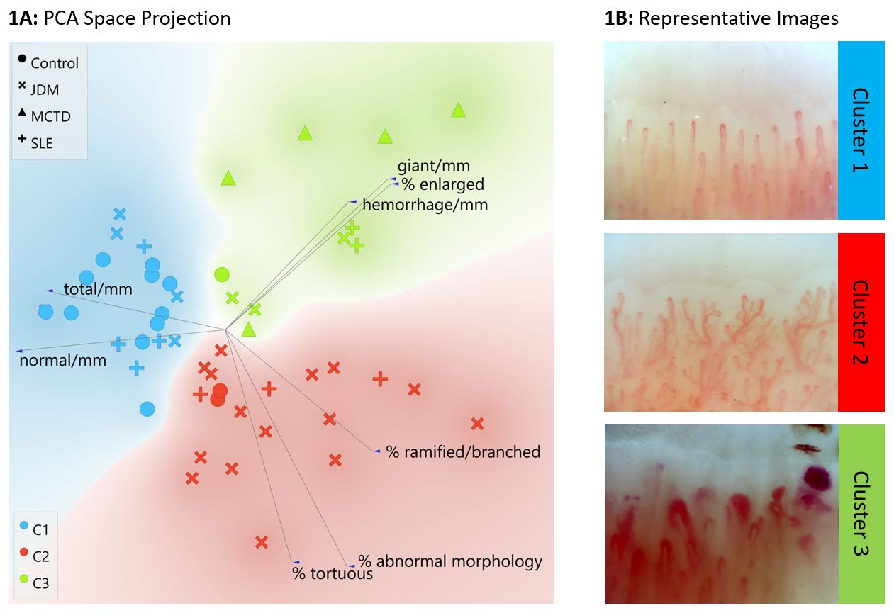
Methods: We utilized nailfold video capillaroscopy at 200x to obtain images from JDM (n=23), childhood systemic lupus erythematosus (SLE) (n=9), mixed connective tissue disease (MCTD) (n=5) and healthy control (n=17) patients at the University of Michigan Pediatric Rheumatology Clinic (1/1/23 – 5/31/23). We quantified NFC features from images using a convolutional neural network via an automated method, including density of normal and giant capillaries, microhemorrhages, and percentage of enlarged, tortuous or ramified capillaries. We then performed unsupervised cluster analysis to identify patients with similar NFC features. Following this, clinical variables were grouped by cluster with differences between means assessed for significance by ANOVA (p≤0.05).

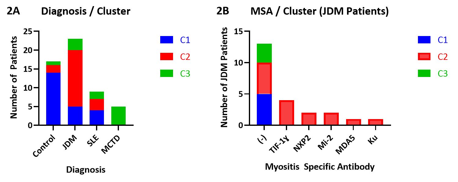
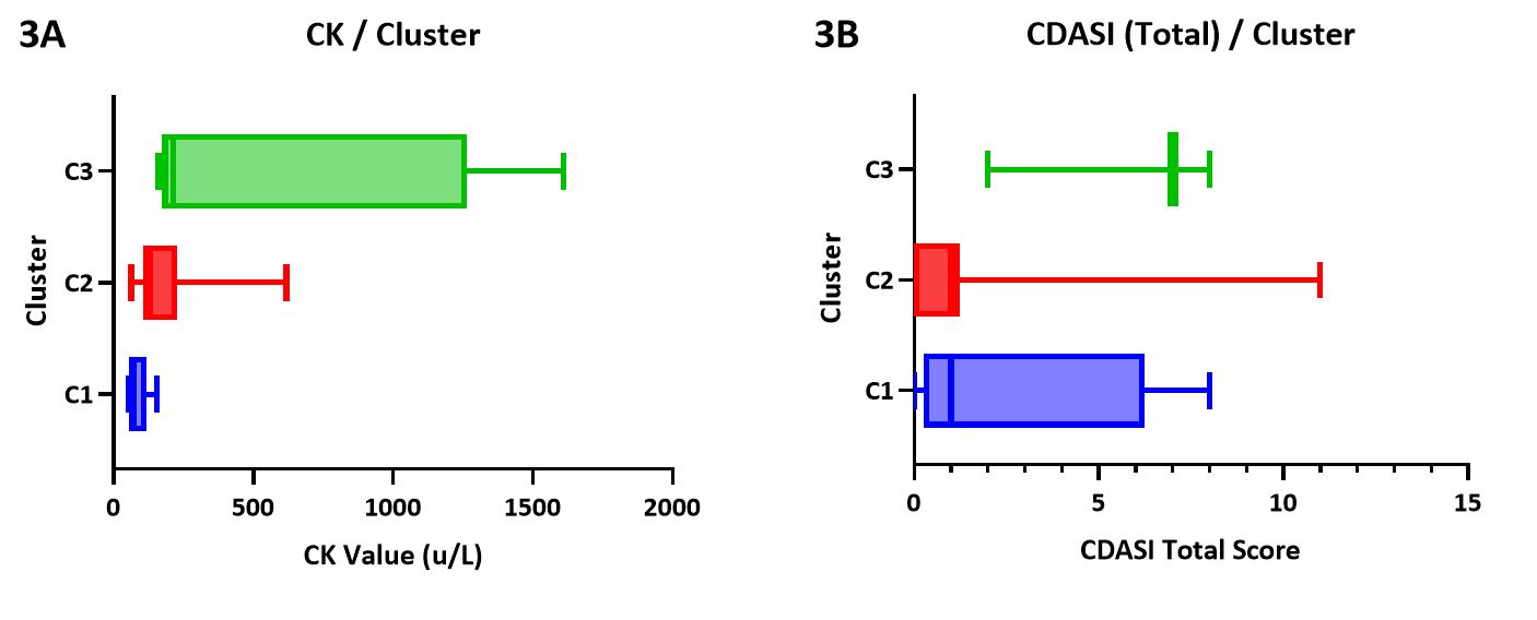
Results: We identified 3 patient clusters distinguished by a predominant NFC pattern of either higher relative capillary density (cluster 1 (C1)), abnormal morphology (C2) or enlarged capillaries (C3) (Figure 1). Upon review of JDM clinical features by cluster, we found JDM patients were primarily assigned to C2 (65%), with the remainder in C1 (22%) and C3 (13%). MCTD patients were entirely assigned to C3 (100%). Patients with SLE were distributed more evenly, with 44%, 33%, and 22% in C1, 2, and 3, respectively. The majority of control patients were assigned to C1 (82%) (Figure 2A). Interestingly, we found that all JDM patients with presence of a positive MSA (TIF-1γ, MDA5, NXP2, Mi-2, Ku) were assigned to C2, while those with negative MSAs were spread between C1, 2 and 3 (Figure 2B). We assessed CK values by cluster, revealing a trend toward higher CK in C3 (mean 550.8 u/L) than C2 (mean 201.2 u/L) or C1 (mean 83.6) (Figure 3A, p=0.073). Aldolase and LDH levels were similar between clusters (p=0.65 and p=0.77, respectively). Skin disease activity as assessed by Cutaneous Dermatomyositis Disease Area and Severity Index (CDASI) scores trended toward higher values in C3 (mean 5.7) as compared to C2 (mean 1.5) and C1 (mean 2.5) (Figure 3B, p=0.124).

Conclusion: We demonstrate 3 distinct groupings of NFC patterns that align with clinical diagnosis and MSA positivity. A predominant pattern of tortuosity and ramification is most pronounced in JDM as compared to MCTD, SLE and control patients. Further investigation into NFC morphology and patterns within a larger JDM cohort may hold the potential to assist in diagnosis, disease subtyping, monitoring and prognostication.

Supporting image 1

Figure 1: (1A) Principal Component Analysis (PCA) plot from Louvain cluster analysis visualized by linear projection. Symbols represent each patient, localized by alignment toward or away from each feature along its axis. (1B) Representative images demonstrating predominant NFC pattern and features from patients in each cluster.

Supporting image 2

Figure 2: Stacked columns representing patients in each assigned cluster, separated by diagnosis (2A, left) and by myositis specific antibody (2B, right).

Supporting image 3

Figure 3: Box-and-whisker plots of CK values by cluster (3A, left) and total CDASI score (3B, right)


Disclosures: N. McClellan: None; S. Vandenbergen: None; S. Matossian: None; J. Kahlenberg: AstraZeneca, 1, Bristol-Myers Squibb(BMS), 2, 5, EMD Serano, 2, exo therapeutics, 2, Gilead, 2, GlaxoSmithKlein(GSK), 1, horizon Therapeutics, 2, Janssen, 5, Pfizer, 2, ROME Therapeutics, 2, 5, Rome Therapeutics, 5, Ventus Therapeutics, 2, 5; J. Turnier: None.

To cite this abstract in AMA style:

McClellan N, Vandenbergen S, Matossian S, Kahlenberg J, Turnier J. Differences in Nailfold Capillary Morphology Distinguish Juvenile Dermatomyositis Patients That Are Myositis-Specific Autoantibody Positive [abstract]. Arthritis Rheumatol. 2023; 75 (suppl 9). https://acrabstracts.org/abstract/differences-in-nailfold-capillary-morphology-distinguish-juvenile-dermatomyositis-patients-that-are-myositis-specific-autoantibody-positive/. Accessed .
  • Tweet
  • Click to email a link to a friend (Opens in new window) Email
  • Click to print (Opens in new window) Print

« Back to ACR Convergence 2023

ACR Meeting Abstracts - https://acrabstracts.org/abstract/differences-in-nailfold-capillary-morphology-distinguish-juvenile-dermatomyositis-patients-that-are-myositis-specific-autoantibody-positive/

Advanced Search

Your Favorites

You can save and print a list of your favorite abstracts during your browser session by clicking the “Favorite” button at the bottom of any abstract. View your favorites »

All abstracts accepted to ACR Convergence are under media embargo once the ACR has notified presenters of their abstract’s acceptance. They may be presented at other meetings or published as manuscripts after this time but should not be discussed in non-scholarly venues or outlets. The following embargo policies are strictly enforced by the ACR.

Accepted abstracts are made available to the public online in advance of the meeting and are published in a special online supplement of our scientific journal, Arthritis & Rheumatology. Information contained in those abstracts may not be released until the abstracts appear online. In an exception to the media embargo, academic institutions, private organizations, and companies with products whose value may be influenced by information contained in an abstract may issue a press release to coincide with the availability of an ACR abstract on the ACR website. However, the ACR continues to require that information that goes beyond that contained in the abstract (e.g., discussion of the abstract done as part of editorial news coverage) is under media embargo until 10:00 AM ET on November 14, 2024. Journalists with access to embargoed information cannot release articles or editorial news coverage before this time. Editorial news coverage is considered original articles/videos developed by employed journalists to report facts, commentary, and subject matter expert quotes in a narrative form using a variety of sources (e.g., research, announcements, press releases, events, etc.).

Violation of this policy may result in the abstract being withdrawn from the meeting and other measures deemed appropriate. Authors are responsible for notifying colleagues, institutions, communications firms, and all other stakeholders related to the development or promotion of the abstract about this policy. If you have questions about the ACR abstract embargo policy, please contact ACR abstracts staff at [email protected].

Wiley

  • Online Journal
  • Privacy Policy
  • Permissions Policies
  • Cookie Preferences

© Copyright 2025 American College of Rheumatology