Session Information
Session Type: Poster Session B
Session Time: 9:00AM-11:00AM
Background/Purpose: There is not cure or an efficient treatment for the osteoarthritis (OA). Mitochondrial damage and dysfunction are described during OA process modulating chondrocyte function and survival contributing to its pathogenesis. As a consequence, mitochondria has proposed as a potential therapeutic target for OA.
Aim: To describe a method to isolate functional mitochondria and assess their functionality and capacity to integrate in joint tissues
Methods: Magnetic beads coupled to anti-TOM22 was used to obtain isolate mitochondria from liver of C57BL/6JOlaHsd after classical tissue digestion. Transmission Electron microscopy (TEM) images and Western blot (WB) were using to stablish the purity and the analysis of Mitotracker Red® staining and ATP were studied in the stablish of mitochondria viability. To evaluated the capacity of mitochondria to integrated in the joint tissues, cartilage and synovial membrane explants were co-incubated in presence of isolated mitochondria labeled with Iron nanoparticles or MitoTarker Red ® during 24, 48 hours (h).Mitochondria labeled with Mitotracker Red® were injected in left knee of 3 C57BL/6JOlaHsd mice and 48 h after the animals were euthanized. The ability of the injected mitochondria to cross the different joint tissues was analyzed, as well as the biological effect and safety of the intra-articular injection.
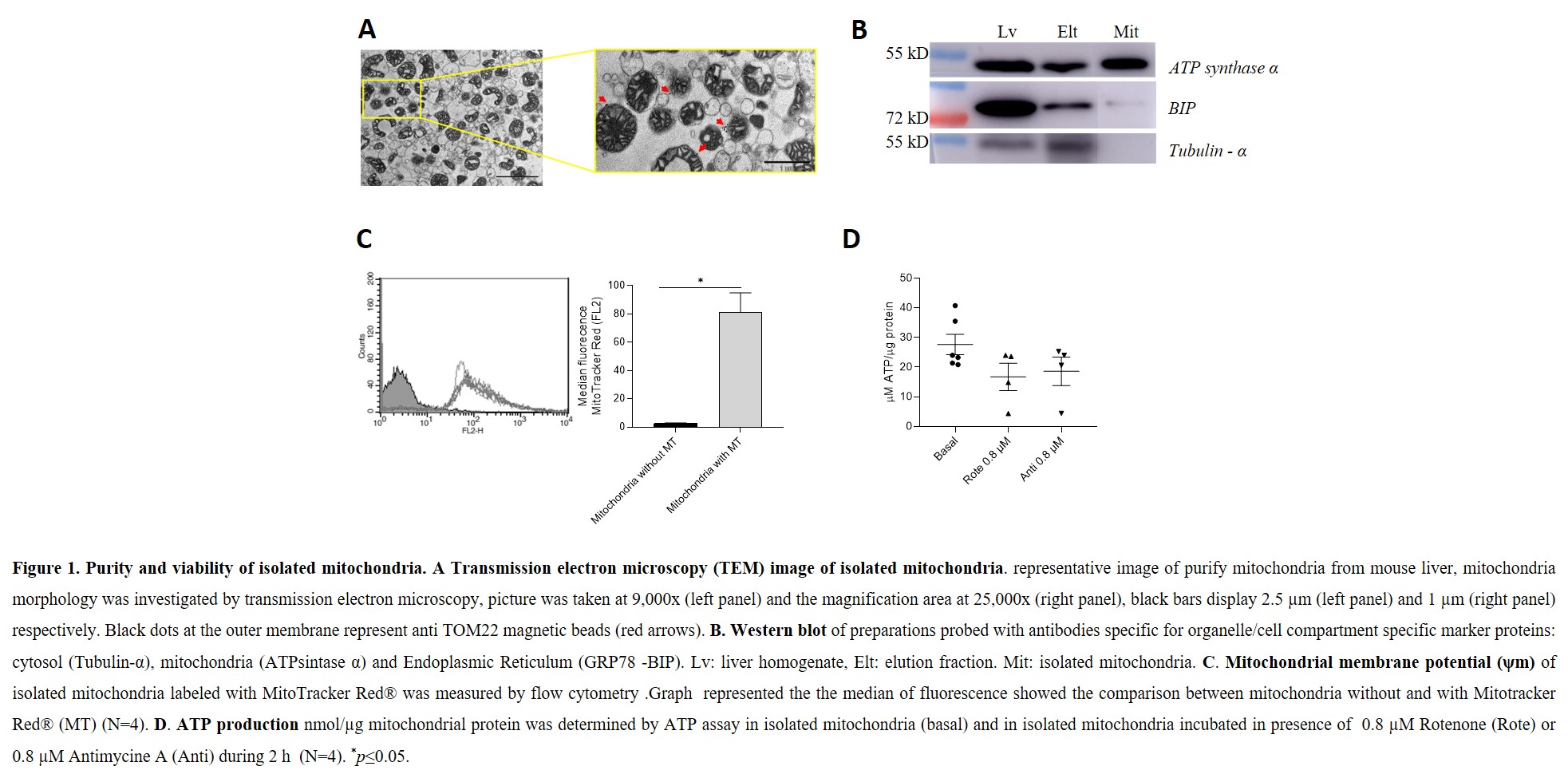
Results: Normal mitochondrial morphology was obtained when TEM images were analyzed (Figure 1-A). WB showed a big signal of ATP synthase α in mitochondrial isolated fraction while BIP and Tubulin-α were detected only in the elution fraction (Figure 1-B). These data reflected that isolated mitochondria were purity. The Mitochondria viability was analyzed through the evaluation of MitoTracker Red ® fluorescence and ATP production. Isolated mitochondria labeled with MitoTracker Red ® showed higher fluorescent than the same samples without label (81±6.92 vs 2.51±0.29, p=0.05) (Figure 1-C). ATP production in isolated mitochondria was modulated when were incubated in presence of 0.8 µM Rotenone (27.64±3.42 vs 16.76±4.62) (Figure 1-D). These results showed that isolated mitochondria were functional.
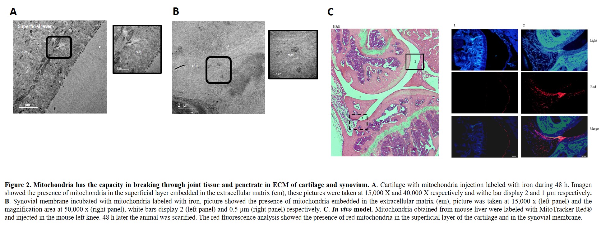
In vitro analysis showed the presence of isolated mitochondria embedded in the ECM of the superficial and intermedial layer of cartilage and into the ECM of synovial tissue (Figure 2-A-B). Morphology of chondrocytes in the different layers of the cartilage and morphology of synoviocytes did not show any change. Intrarticular injection of mitochondria caused low grade of joint inflammatory response as well as any systemic complication, no animal dead. Histologic studies of left joint showed Mitotracker Red® signal in the synovial membrane and in the superficial layer of the cartilage (Figure 2-C). Synovitis was not present. Histologic studies of the right knee and organs did not detect any red fluorescence stain.
Conclusion: Isolated mitochondria penetrate ECM of cartilage and synovium. In mice, intraarticular injection of functional isolated mitochondria is safety. These data give us the opportunity to continue studying the mitochondria as a treatment for OA.
To cite this abstract in AMA style:
Fernandez-Moreno M, Hermida-Gomez T, Paniagua-Barro S, Rodriguez-Coello A, Vaamonde-garcia C, Blanco F. Development of a Method to Isolate Functional Mitochondria and Assess Their Functionality and Integration in Joint Tissues: In Vitro and in Vivo Models [abstract]. Arthritis Rheumatol. 2023; 75 (suppl 9). https://acrabstracts.org/abstract/development-of-a-method-to-isolate-functional-mitochondria-and-assess-their-functionality-and-integration-in-joint-tissues-in-vitro-and-in-vivo-models/. Accessed .« Back to ACR Convergence 2023
ACR Meeting Abstracts - https://acrabstracts.org/abstract/development-of-a-method-to-isolate-functional-mitochondria-and-assess-their-functionality-and-integration-in-joint-tissues-in-vitro-and-in-vivo-models/


