Session Information
Date: Sunday, November 8, 2020
Session Type: Poster Session C
Session Time: 9:00AM-11:00AM
Background/Purpose: Fundoscopy is essential to identify the retinopathy of patients with autoimmune diseases. However, the classification of different autoimmune diseases is difficult by human eyes using traditional fundoscopy. We hypothesized that patients with axial spondyloarthritis (SpA) might have subtle differences in retinal images that are sufficient to classify SpA. We aimed to develop a deep learning model to detect the difference in retinal images between patients with SpA and other autoimmune diseases.
Methods: We prospectively collected 342 retinal images from 218 patients with or without SpA, which diagnosis was based on the Assessment of SpondyloArthritis international Society classification criteria for axial spondyloarthritis. The population was divided into a training group, a validation group and testing group (5:1:1). The retinal images were fed to a model based on the EfficientNet-B1, which was trained to classify SpA. The performance of the model was tested on a hold-out dataset of 31 patients whose retinal images were not used in the model training. The essential pixels that the models used to classify SpA was visualized using the Gradient-weighted Class Activation Mapping (Grad-CAM). Overall model performance was measured by accuracy, sensitivity, specificity, area under the curve of the receiver operator curve (AUROC).
Results: We included 51 women and 93 men with SpA (mean age, 42.9±12.8 years) and 74 non-SpA individuals (F: M, 57:17; mean age, 49.4±13 years). Among them, images from 187 patients were used for training and validation, while 31 of them were used to test the model performance. The overall model performance was 0.735 by AUROC. The sensitivity and specificity to classify SpA was 87% and 62.5%, respectively. Figures 1 and 2 showed the results from Grad-CAM, which highlighted the model interpretability.
Conclusion: This exploratory study indicates that subtle changes in retinal images may be distinctive for SpA which may be detected by deep learning models.
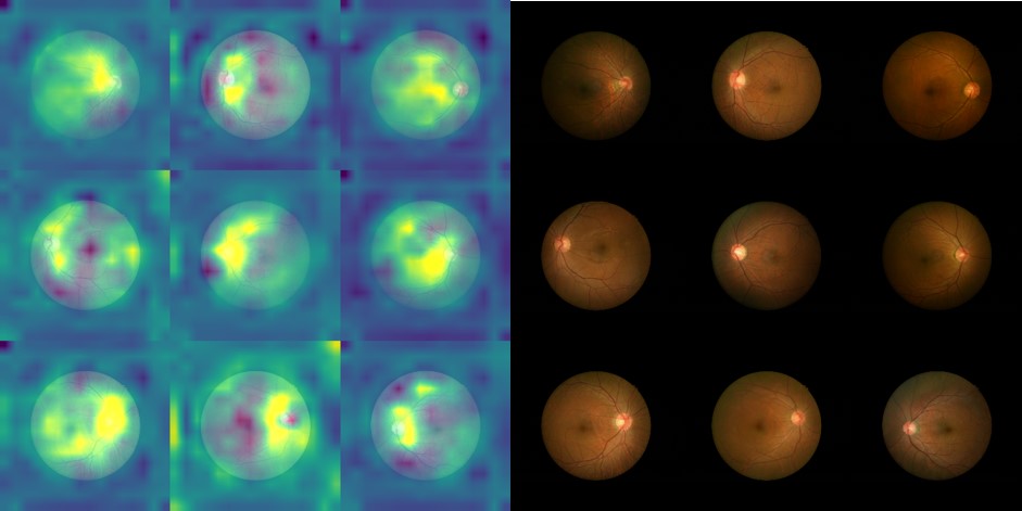 Highlighting important regions of fundoscopy localized by Grad-CAM(left) and original fundoscopy(right) in SpA patients.
 Highlighting important regions of fundoscopy localized by Grad-CAM(left) and original fundoscopy(right) in non-SpA patients.
To cite this abstract in AMA style:
Huang Y, Kuo C, Huang Y, Hwang Y, Lin C. Detecting Subtle Changes in Fundoscopic Retinal Images in Patients with Axial Spondyloarthritis with Deep Learning [abstract]. Arthritis Rheumatol. 2020; 72 (suppl 10). https://acrabstracts.org/abstract/detecting-subtle-changes-in-fundoscopic-retinal-images-in-patients-with-axial-spondyloarthritis-with-deep-learning/. Accessed .« Back to ACR Convergence 2020
ACR Meeting Abstracts - https://acrabstracts.org/abstract/detecting-subtle-changes-in-fundoscopic-retinal-images-in-patients-with-axial-spondyloarthritis-with-deep-learning/