Session Information
Date: Monday, November 9, 2015
Title: Imaging of Rheumatic Diseases Poster II: X-ray, MRI, PET and CT
Session Type: ACR Poster Session B
Session Time: 9:00AM-11:00AM
Background/Purpose: Radiographs have been traditionally used
to detect bone erosions and joint space narrowing (JSN) in rheumatoid arthritis
(RA), but they are not sensitive to small erosions and subtle changes in
erosions or JSN based on Sharp scoring, which requires at least 12 months to
detect disease progression in clinical trials1. High-resolution
peripheral quantitative computed tomography (HR-pQCT) is an emerging in vivo imaging technology that has an in vivo resolution 82 μm that allows us to detect smaller erosions2,
measure joint space width (JSW), quantify trabecular bone structure, and detect
subtle changes in these measures. In this study, we examine the effects of
anti-TNF on JSW and bone structure in a three-month period.
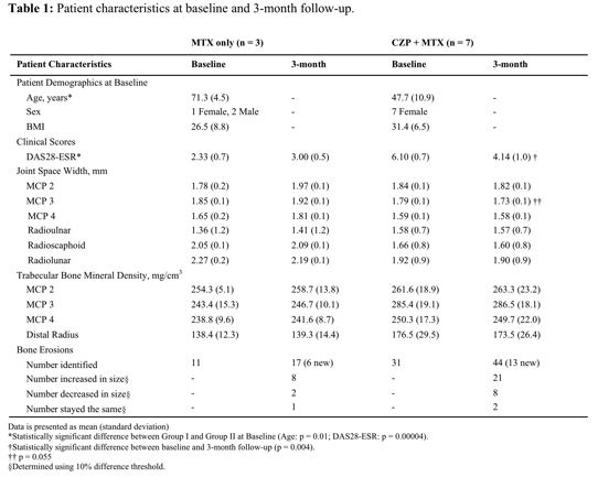
Methods: The wrists of 10 RA patients (54.8 ± 15.1 year.,
8 female., DAS28-ESR: 4.97 ± 1.83) were scanned using HR-pQCT with an
isotropic resolution of 82 μm. Patients
were divided into two groups: Group I was given only methotrexate (MTX) (3
patients) and Group II was given certolizumab pegol (CZP) and MTX (7 patients).
Scans were performed on metacarpophalangeal (MCP), and wrist joints at baseline
(before anti-TNF initiation for Group II) and 3-month follow up. The second,
third, and fourth MCP, and wrist JSW were measured, and the MCP and distal
radius trabecular bone were quantified using previously developed methods3.
Bone erosion dimensions were evaluated by measuring the maximal erosion
dimensions on an axial 2-dimensional slice4. T-tests were performed
to compare measures between the groups and within the groups from baseline to
3-month.
Results: DAS28-ESR of patients in Group II
decreased significantly from baseline to 3-month (p = 0.004), and the decrease
in MCP 3 JSW for Group II approached significance (p = 0.055). Eleven and 31
erosions were identified at baseline for Group I and II respectively, with
72.7% and 67.7% of these lesions increasing in size (change > 10%) at
baseline and 3-month respectively. Six and 13 new erosions were identified at
3-month for Group I and II respectively.
Conclusion: The decrease of MCP 3 JSW in Group II may
be due to decreased inflammation and less synovitis and effusion with anti-TNF
treatment. We observed more than half of the erosions increased in size and the
generation of new erosions for both groups within a very short time period (3
months) using HR-pQCT, indicating the high sensitivity of HR-pQCT detecting
subtle changes in erosions as compared to radiographs. The significant decrease
in DAS28-ESR for Group II suggests that although anti-TNF treatment suppresses
the inflammation pathway, the process of structural joint damage may require more
time to be arrested.
[1] Heijde et al,
The Journal of Rheumatology. 1995. [2] Lee et al, International Journal of
Rheumatic Diseases. 2013 [3] Yang et al, International Journal of Rheumatic
Diseases. 2015 [4] Srikhum et al, The Journal of Rheumatology. 2013
To cite this abstract in AMA style:
Tanaka M, Su F, Lee MH, Burghardt AJ, Link TM, Graf JD, Imboden J, Li X. Detecting Subtle Changes in Bone Structure Following Three-Month Anti-TNF Theraphy Using High-Resolution Peripheral Quantitative Computed Tomography [abstract]. Arthritis Rheumatol. 2015; 67 (suppl 10). https://acrabstracts.org/abstract/detecting-subtle-changes-in-bone-structure-following-three-month-anti-tnf-theraphy-using-high-resolution-peripheral-quantitative-computed-tomography/. Accessed .« Back to 2015 ACR/ARHP Annual Meeting
ACR Meeting Abstracts - https://acrabstracts.org/abstract/detecting-subtle-changes-in-bone-structure-following-three-month-anti-tnf-theraphy-using-high-resolution-peripheral-quantitative-computed-tomography/

