ACR Meeting Abstracts

ACR Meeting Abstracts

  • Meetings
    • ACR Convergence 2024
    • ACR Convergence 2023
    • 2023 ACR/ARP PRSYM
    • ACR Convergence 2022
    • ACR Convergence 2021
    • ACR Convergence 2020
    • 2020 ACR/ARP PRSYM
    • 2019 ACR/ARP Annual Meeting
    • 2018-2009 Meetings
    • Download Abstracts
  • Keyword Index
  • Advanced Search
  • Your Favorites
    • Favorites
    • Login
    • View and print all favorites
    • Clear all your favorites
  • ACR Meetings

Abstract Number: 0059

Deep Immune Cell Profiling of Tissue Microenvironment Using Imaging Mass Cytometry in Psoriatic Disease

Lihi Eder1, Stephane Caucheteux1, Somi Afiuni2, Adriana Krizova1, James Limacher1, Hartland Jackson1 and Vincent Piguet1, 1University of Toronto, Toronto, ON, Canada, 2Lunenfeld Tanenbaum Research Institute, Sinai Health System, Toronto, ON, Canada

Meeting: ACR Convergence 2021

Keywords: immunology, innate immunity, Psoriatic arthritis, spondyloarthritis

  • Tweet
  • Click to email a link to a friend (Opens in new window) Email
  • Click to print (Opens in new window) Print
Session Information

Date: Saturday, November 6, 2021

Title: Spondyloarthritis Including PsA – Basic Science Poster (0046–0068)

Session Type: Poster Session A

Session Time: 8:30AM-10:30AM

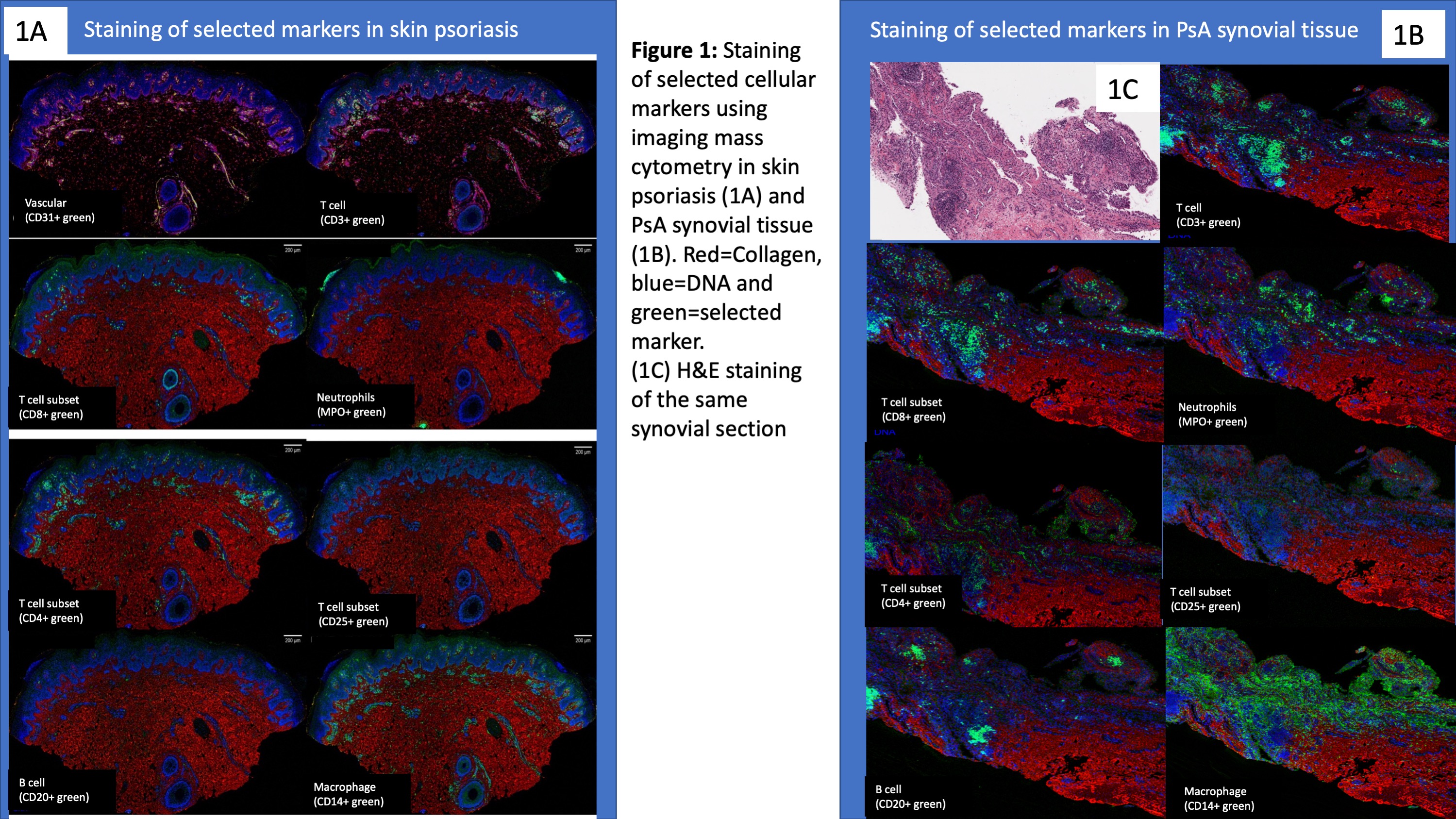
Background/Purpose: Cross talk between different cell types, such as T cell subsets and other immune cell populations, is important in psoriatic disease. Imaging Mass Cytometry (IMC) is a novel technology that enables comprehensive analysis of cellular phenotypes and their interrelationships at the tissue level. We performed a multi-parameter characterization of various immune cell populations and assessed their spatial interactions in skin and synovial tissue samples from patients with psoriatic disease.

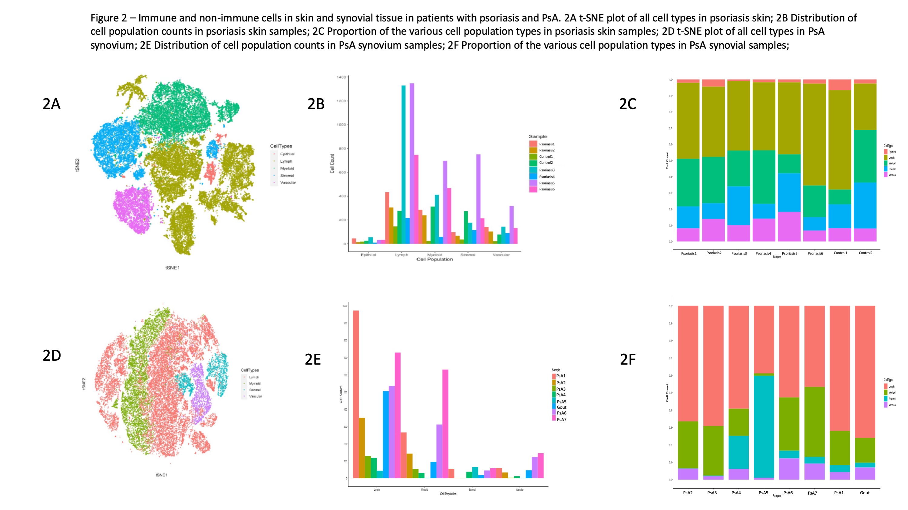
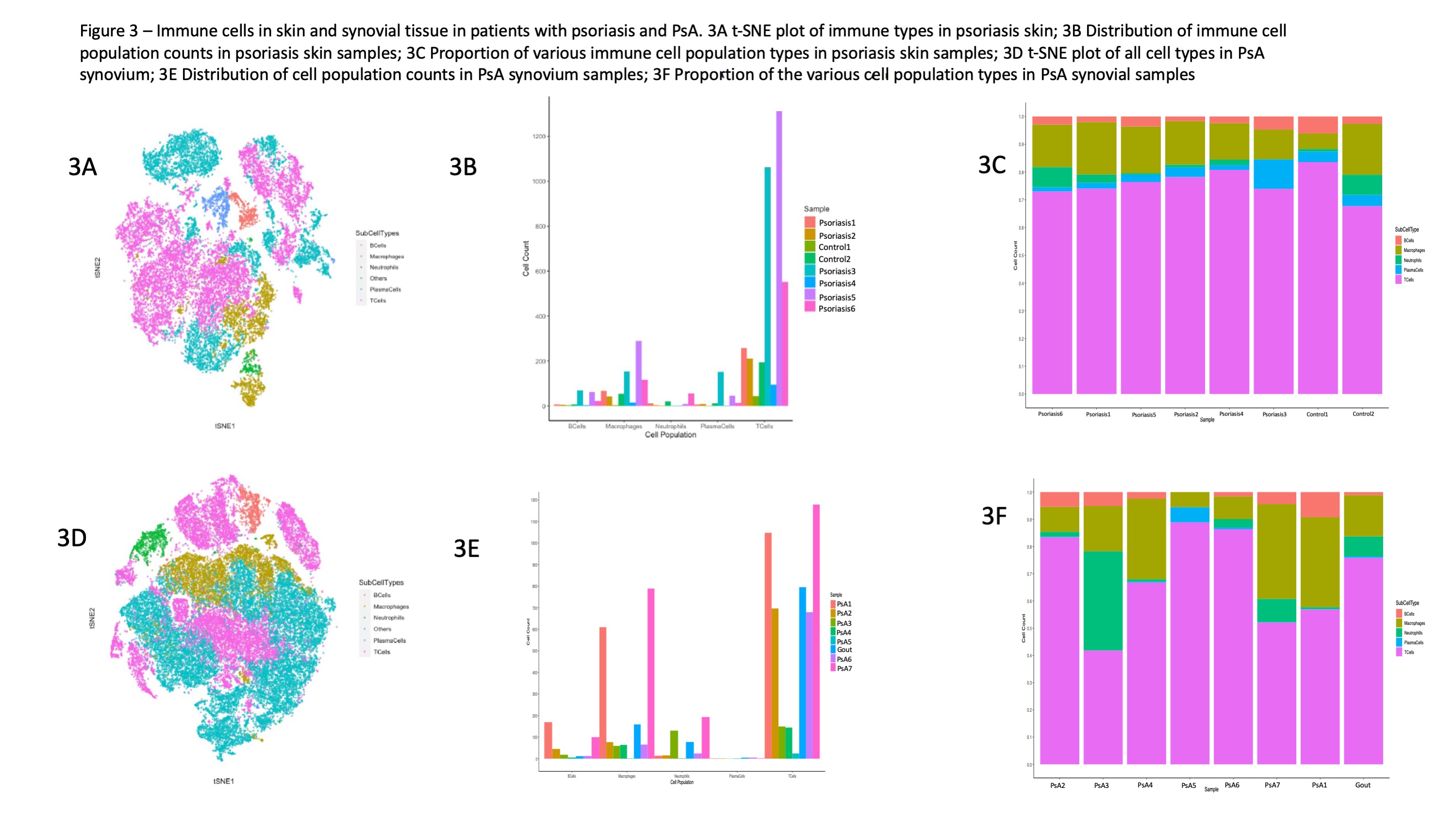
Methods: A total of 6 skin and 7 synovial formalin-fixed, paraffin embedded samples from patients with plaque psoriasis and psoriatic arthritis (PsA), respectively, were analyzed. Additionally, 1 synovial sample from a gout patients and a skin sample from a control patient were analyzed. We used a panel of 36 of metal-tagged antibodies to stain different cell populations. IMC was performed using the Hyperion Imaging System (Fluidigm). Tissue of 5-µm thickness was ablated with Nd:YAG 213 mm laser and analyzed in a Fluidigm CyTOF mass cytometer. Images were analyzed using MCD Viewer. Data were converted to TIFF format and segmented into single cells using previously developed methods. Individual cells were segmented using a combination of in-house program and CellProfiler to classify pixels on the basis of a combination of antibody stains to identify membranes and nuclei. The maps were then segmented into single-cell object masks using CellProfiler. Cell were classified into cell categories based on pre-specified markers (epithelial, vascular, stromal, myeloid and lymphoid) and further classified into immune cells types (T cell, macrophage, B cell, neutrophils, plasma cells). Single-cell marker expressions were summarized by mean pixel values for each channel. To visualize the number of cells per image the cell counts were normalized by the image area (total number of pixels) and displayed as cell density.

Results: Markers identifying stromal and immune cell types performed well and correlated with the expected location and morphological features of the various cell types (Figure 1). Among all cell types in the skin and synovium, lymphoid cells accounted for the most prevalent cell type followed by myeloid cells (Figure 2A-2B). Large degree of inter-patient heterogeneity was found in the prevalence of the various immune cell population across the different skin and synovial samples (Figure 3A-3B). T cells accounted for the largest immune cell type in both skin and synovial samples followed by macrophages and lower counts of neutrophils, B cells and plasma cells. Neighborhood analysis showed high correlation between CD20+, CD3+, CD4+ and CD8+ in the synovial tissue, which suggest close physical proximity and cross-talk between these T cell subsets and B cells in the synovium.

Conclusion: Innate and adaptive immune cells can be reliably identified using IMC in skin and synovial tissue. Cross talk between T cell subsets and innate immune cells in the skin and synovial tissue plays a role in psoriatic disease. IMC technology provides opportunities for exploring the underlying mechanisms driving psoriasis and PsA.

Figure 1: Staining of selected cellular markers using imaging mass cytometry in skin psoriasis (1A) and PsA synovial tissue (1B). Red=Collagen, blue=DNA and green=selected marker. (1C). H&E staining of the same synovial section

Figure 2 – Immune and non-immune cells in skin and synovial tissue in patients with psoriasis and PsA. 2A t-SNE plot of all cell types in psoriasis skin; 2B Distribution of cell population counts in psoriasis skin samples; 2C Proportion of the various cell population types in psoriasis skin samples; 2D t-SNE plot of all cell types in PsA synovium; 2E Distribution of cell population counts in PsA synovium samples; 2F Proportion of the various cell population types in PsA synovial samples

Figure 3 – Immune cells in skin and synovial tissue in patients with psoriasis and PsA. 3A t-SNE plot of immune types in psoriasis skin; 3B Distribution of immune cell population counts in psoriasis skin samples; 3C Proportion of various immune cell population types in psoriasis skin samples; 3D t-SNE plot of all cell types in PsA synovium; 3E Distribution of cell population counts in PsA synovium samples; 3F Proportion of the various cell population types in PsA synovial samples


Disclosures: L. Eder, Pfizer, 1, 5, UCB, 5, Abbvie, 1, 5, Novartis, 2, Eli Lily, 1, 5, Fluidigm, 5, 12, Family member - employee, Janssen, 5; S. Caucheteux, None; S. Afiuni, None; A. Krizova, None; J. Limacher, Sanofi Genzyme, 2; H. Jackson, GlaxoSmithKline, 7, Fluidigm, 6, Abcam, 12, Collaboration; V. Piguet, None.

To cite this abstract in AMA style:

Eder L, Caucheteux S, Afiuni S, Krizova A, Limacher J, Jackson H, Piguet V. Deep Immune Cell Profiling of Tissue Microenvironment Using Imaging Mass Cytometry in Psoriatic Disease [abstract]. Arthritis Rheumatol. 2021; 73 (suppl 9). https://acrabstracts.org/abstract/deep-immune-cell-profiling-of-tissue-microenvironment-using-imaging-mass-cytometry-in-psoriatic-disease/. Accessed .
  • Tweet
  • Click to email a link to a friend (Opens in new window) Email
  • Click to print (Opens in new window) Print

« Back to ACR Convergence 2021

ACR Meeting Abstracts - https://acrabstracts.org/abstract/deep-immune-cell-profiling-of-tissue-microenvironment-using-imaging-mass-cytometry-in-psoriatic-disease/

Advanced Search

Your Favorites

You can save and print a list of your favorite abstracts during your browser session by clicking the “Favorite” button at the bottom of any abstract. View your favorites »

All abstracts accepted to ACR Convergence are under media embargo once the ACR has notified presenters of their abstract’s acceptance. They may be presented at other meetings or published as manuscripts after this time but should not be discussed in non-scholarly venues or outlets. The following embargo policies are strictly enforced by the ACR.

Accepted abstracts are made available to the public online in advance of the meeting and are published in a special online supplement of our scientific journal, Arthritis & Rheumatology. Information contained in those abstracts may not be released until the abstracts appear online. In an exception to the media embargo, academic institutions, private organizations, and companies with products whose value may be influenced by information contained in an abstract may issue a press release to coincide with the availability of an ACR abstract on the ACR website. However, the ACR continues to require that information that goes beyond that contained in the abstract (e.g., discussion of the abstract done as part of editorial news coverage) is under media embargo until 10:00 AM ET on November 14, 2024. Journalists with access to embargoed information cannot release articles or editorial news coverage before this time. Editorial news coverage is considered original articles/videos developed by employed journalists to report facts, commentary, and subject matter expert quotes in a narrative form using a variety of sources (e.g., research, announcements, press releases, events, etc.).

Violation of this policy may result in the abstract being withdrawn from the meeting and other measures deemed appropriate. Authors are responsible for notifying colleagues, institutions, communications firms, and all other stakeholders related to the development or promotion of the abstract about this policy. If you have questions about the ACR abstract embargo policy, please contact ACR abstracts staff at [email protected].

Wiley

  • Online Journal
  • Privacy Policy
  • Permissions Policies
  • Cookie Preferences

© Copyright 2025 American College of Rheumatology