ACR Meeting Abstracts

ACR Meeting Abstracts

  • Meetings
    • ACR Convergence 2024
    • ACR Convergence 2023
    • 2023 ACR/ARP PRSYM
    • ACR Convergence 2022
    • ACR Convergence 2021
    • ACR Convergence 2020
    • 2020 ACR/ARP PRSYM
    • 2019 ACR/ARP Annual Meeting
    • 2018-2009 Meetings
    • Download Abstracts
  • Keyword Index
  • Advanced Search
  • Your Favorites
    • Favorites
    • Login
    • View and print all favorites
    • Clear all your favorites
  • ACR Meetings

Abstract Number: 0869

Deciphering Pathogenic Phenotypes by Multi-modal Deep Single-cell Blood Immunophenotyping in Individuals At-risk for Rheumatoid Arthritis

Jun Inamo1, Joshua Keegan2, Alec Griffith2, Tusharkanti Ghosh1, Alice Horisberger2, Kaitlyn Howard2, John Pulford2, Ekaterina Murzin2, Brandon Hancock2, Thomas Eisenhaure3, Salina Dominguez4, Miranda Gurra5, Siddarth Gurajala3, Anna Helena Jonsson1, Jennifer Seifert6, Marie Feser7, Jill Norris8, Ye Cao2, William Apruzzese9, S. Louis Bridges10, Vivian Bykerk11, Susan Goodman12, Laura Donlin11, Gary S. Firestein13, Joan Bathon14, Laura B. Hughes15, Darren Tabechian16, Andrew Filer17, Costantino Pitzalis18, Jennifer Anolik19, Larry Moreland20, Nir Hacohen21, Joel Guthridge22, Judith James22, Carla Cuda5, Harris Perlman5, Michael B. Brenner2, Soumya Raychaudhuri23, Jeffrey Sparks24, Michael Holers7, Kevin Deane25, James A. Lederer26, Deepak Rao26 and Fan Zhang27, and the Accelerating Medicines Partnership RA/SLE Network, 1University of Colorado School of Medicine, Aurora, CO, 2Brigham and Women's Hospital and Harvard Medical School, Boston, MA, 3Broad Institute of MIT and Harvard, Cambridge, 4Northwestern University, Chicago, 5Northwestern University, Chicago, IL, 6University of Colorado and Oklahoma Medical Research Foundation, Aurora, CO, 7Division of Rheumatology, University of Colorado School of Medicine, Aurora, CO, 8Colorado School of Public Health, Denver, CO, 9Accelerating Medicines Partnership® Program: Rheumatoid Arthritis and Systemic Lupus Erythematosus (AMP® RA/SLE) Network, Boston, MA, 10Division of Rheumatology, Weill Cornell Medical College, New York, NY, 11Hospital For Special Surgery, New York, NY, 12Hospital for Special Surgery, New York 10025, NY, 13University of California, San Diego, La Jolla, 14Columbia University, New York, NY, 15University of Alabama at Birmingham Medicine, Birmingham, AL, 16University of Rochester Medical Center, Rochester, 17Rheumatology Research Group, Institute for Inflammation and Ageing, NIHR Birmingham Biomedical Research Center and Clinical Research Facility, University of Birmingham, Birmingham, United Kingdom, 18QMUL, Bromley Kent, United Kingdom, 19University of Rochester Medical Center, Rochester, NY, 20University of Colorado, Denver, CO, 21Broad Institute of MIT and Harvard, Boston, MA, 22Oklahoma Medical Research Foundation, Oklahoma City, OK, 23Brigham and Women's Hospital, Boston, MA, 24Brigham and Women's Hospital and Harvard Medical School, Boston, MA, USA, Boston, MA, 25University of Colorado Denver Anschutz Medical Campus, Aurora, CO, 26Brigham and Women's Hospital, Harvard Medical School, Boston, MA, 27University of Colorado, Aurora, CO

Meeting: ACR Convergence 2024

Keywords: Bioinformatics, genomics, rheumatoid arthritis, T Cell

  • Tweet
  • Email
  • Print
Session Information

Date: Saturday, November 16, 2024

Title: Abstracts: RA – Diagnosis, Manifestations, & Outcomes II: Bad Blood (Serologic and Imaging Biomarkers)

Session Type: Abstract Session

Session Time: 3:00PM-4:30PM

Background/Purpose: Rheumatoid arthritis (RA) is a systemic autoimmune disease with currently no effective prevention strategies. Single-cell technologies have been recently used to investigate established RA heterogeneity, but it is unknown if the immune populations identified from RA tissues are playing important roles in blood during the preclinical phase of disease. Thus, identifying pathogenic immune phenotypes in individuals who can be at risk for future RA, “At-Risk RA”, is crucial to establishing prevention strategies.

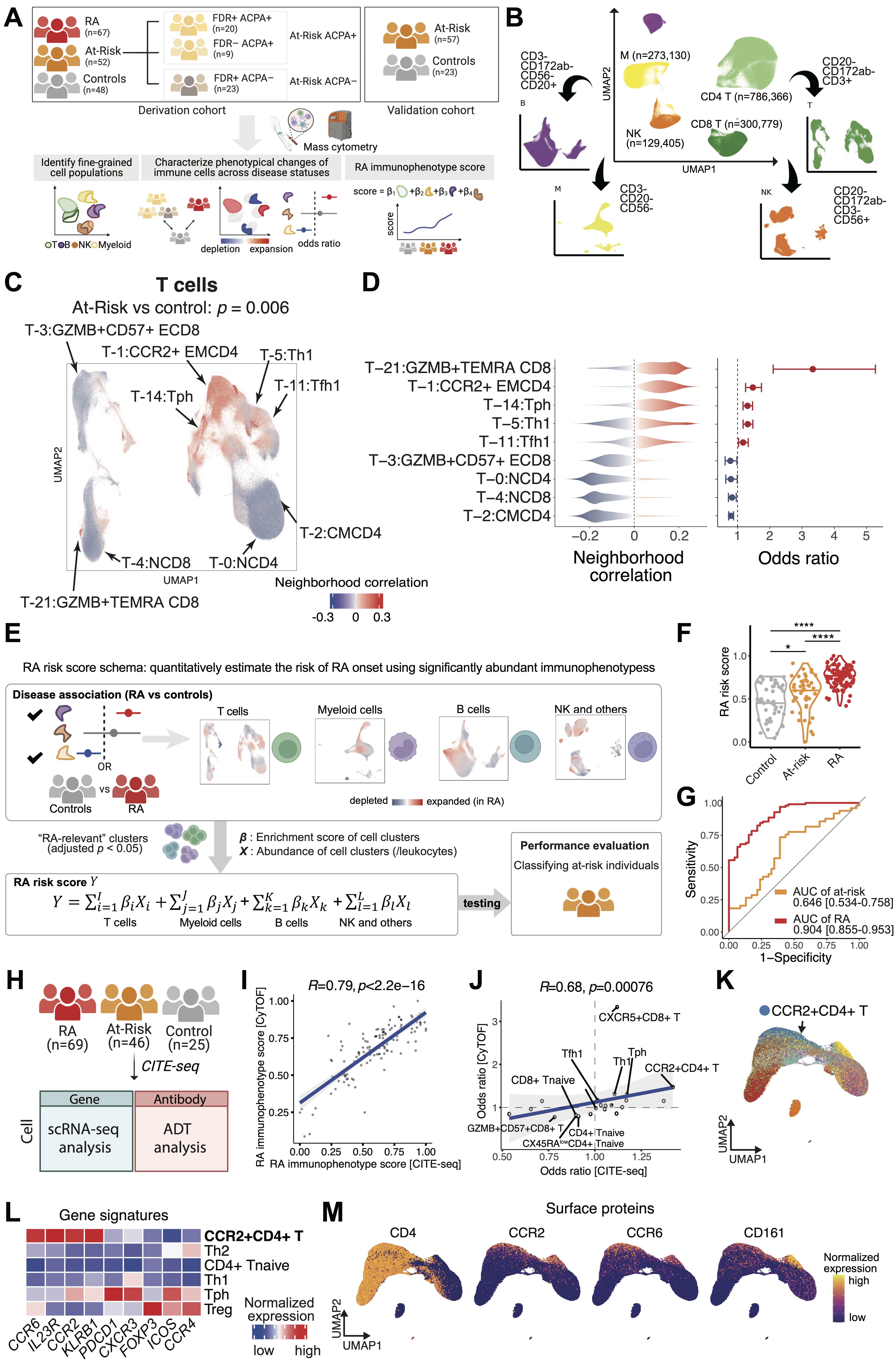
Methods: We applied scalable computational strategies on mass cytometry data to deeply characterize immunophenotypes in blood from At-Risk individuals of clinical subpopulations based on antibodies to citrullinated protein antigens (ACPA) and first-degree relative (FDR) (n=52), and from established RA (n=67), and healthy controls (n=48) (Figure 1A-B). Further, we employed CITE-seq data to blood from At-Risk individuals (n=46), established RA (n=69), and healthy controls (n=25) to validate our findings.

Results: Through integrative and disease association analyses, we quantified the immune populations and uncovered significant cell expansions in At-Risk individuals compared with controls, including CCR2+ T helper cells, T peripheral helper cells (Tphs), type 1 T helper cells, and GZMB+ effector memory T cells that re-express CD45RA (TEMRA) cytotoxic T cells (Figure 1C-D). We further validated the At-Risk associations of these T cell phenotypes using our validation cohort of 57 At-Risk and 23 healthy individuals. In addition, we found that CD15+ classical monocytes were highly expanded in ACPA-negative At-Risk, and an activated PAX5low naïve B cell population was expanded in ACPA-positive individuals who also had an FDR with RA. Further, we developed a “RA immunophenotype score” classification method based on the degree of enrichment and the abundance of cell states relevant to established RA using mixed-effect modeling and logistic regression (Figure 1E). We found this score significantly distinguished At-Risk individuals from the controls (p=0.039 and AUC >0.6) (Figure 1F-G). In CITE-seq data (Figure 1H), we observed significant correlation of both RA immunophenotype scores (Figure 1I) and At-Risk-association effect size of T cell subsets (Figure 1J) between those derived from CyTOF data and those obtained from CITE-seq data, reinforcing the robustness and reliability of our scoring method across diverse technological platforms. Finally, we discovered high expression of Th17-related genes in CCR2+CD4+ T cells in CITE-seq data (Figure 1K-M).

Conclusion: We systematically characterized altered circulating immune phenotypes in At-Risk individuals, and  immunophenotypical differences among ACPA+ and FDR At-Risk subpopulations. Our classification model may provide a promising approach to understand the pathogenesis of RA with the goal to developing preventive strategies.

Supporting image 1

Figure 1: Overview of mass cytometry analytical strategy, clustering, and classifications for At-Risk RA and established RA individuals. A. Description of study design regarding patient recruitment, clinical classification, and computational strategies. B. Gating strategy for mass cytometry data to determine selected immune cell populations. C. Identifications of specific T cell populations that were associated with At-Risk. Cells in UMAP are colored in red (expansion) or blue (depletion) and p-value is shown as well. D. Distributions of cell neighborhood correlations and odds ratios. Error bars for odds ratio represent 95% confidence intervals. E. RA immunophenotype score utilizing RA-specific cell type abundances to quantify and distinguish At-Risk individuals from control. For each cell type, all p-values from the covarying neighborhood analysis test were p = 1e_3. We incorporated clusters that are significantly associated with RA (adjusted p < 0.05) to model the RA immunophenotype score. We calculated RA immunophenotype score based on cell type abundances multiplied by corresponding major cell type proportions and enrichment scores for each cell type, F. Distribution of RA immunophenotype score across individual samples from RA, At-Risk, and controls; **** p < 0.0001, * p < 0.05, G. Receiver operating characteristic (ROC) analysis to evaluate the classification performance of RA immunophenotype score in distinguishing At-Risk from control. Areas under the curve (AUC) with 95% confidence intervals were described. All the analyses are adjusted for age and sex, H. Experimental design of the CITE-seq data, I. Scatter plot displaying the correlation between RA immunophenotype scores for overlapping individuals (n=124) derived from CyTOF data and CITE-seq data, J. Scatter plot showing the correlation between odds ratios for At-Risk association for various T cell clusters. Significantly associated clusters in the CyTOF analysis are labeled, K. UMAP plot of T cells from CITE-seq data. CCR2+ CD4+ T cells are labeled and colored in blue, L, Heatmap showing normalized expression levels of Th17-related genes across different helper T cell subsets, M. UMAP plots depicting the expression patterns of Th17-related surface proteins.


Disclosures: J. Inamo: None; J. Keegan: None; A. Griffith: None; T. Ghosh: None; A. Horisberger: None; K. Howard: None; J. Pulford: None; E. Murzin: None; B. Hancock: None; T. Eisenhaure: None; S. Dominguez: None; M. Gurra: None; S. Gurajala: None; A. Jonsson: Pfizer, 6; J. Seifert: None; M. Feser: None; J. Norris: None; Y. Cao: None; W. Apruzzese: Pfizer, 3; S. Bridges: None; V. Bykerk: BMS, 5, Pfizer, 1; S. Goodman: Novartis Corporation Pharmaceuticals, 5, UCB, 1; L. Donlin: Bristol-Myers Squibb(BMS), 2, Karius, Inc., 5, Stryker, 2; G. Firestein: Eli Lilly, 5; J. Bathon: None; L. Hughes: None; D. Tabechian: Amgen, 12, share holder; A. Filer: None; C. Pitzalis: AbbVie/Abbott, 2, 5, 6, AnaptysBio, 2, 5, Exagen, 2, Janssen, 2, 5, 6, Kinikska, 2, Novartis, 2, 5, Pfizer, 5, Sanofi, 2, 5, 6; J. Anolik: None; L. Moreland: None; N. Hacohen: None; J. Guthridge: AstraZeneca, 5, Bristol-Myers Squibb(BMS), 5; J. James: GlaxoSmithKlein(GSK), 1, Progentec Diagnostics, Inc., 5, 10; C. Cuda: None; H. Perlman: Abbvie, 2, AnaptysBio, 12, Speaking, advising, consulting, or providing educational programs, Exagen, 2, Janssen, 2, Kiniksa, 2; M. Brenner: GlaxoSmithKlein(GSK), 2, Mestag Therapeutics, 2, 11, Moderna, 2; S. Raychaudhuri: Janssen, 1, Mestag, 8, Nimbus, 2, Pfizer, 1, Sonoma, 8, Third Rock Ventures, 2; J. Sparks: Boehringer-Ingelheim, 2, 5, Bristol-Myers Squibb(BMS), 2, 5, Gilead, 2, Janssen, 2, Pfizer, 2, UCB, 2; M. Holers: None; K. Deane: Boehringer-Ingelheim, 5, Bristol-Myers Squibb(BMS), 6, Gilead, 5, Inova, 6, 12, Material Support, ThermoFisher, 5, 6; J. Lederer: None; D. Rao: Amgen, 6, AnaptysBio, 2, AstraZeneca, 1, Bristol-Myers Squibb, 2, 5, GlaxoSmithKline, 2, HiFiBio, 2, Janssen, 5, Merck, 5, Scipher Medicine, 2; F. Zhang: None.

To cite this abstract in AMA style:

Inamo J, Keegan J, Griffith A, Ghosh T, Horisberger A, Howard K, Pulford J, Murzin E, Hancock B, Eisenhaure T, Dominguez S, Gurra M, Gurajala S, Jonsson A, Seifert J, Feser M, Norris J, Cao Y, Apruzzese W, Bridges S, Bykerk V, Goodman S, Donlin L, Firestein G, Bathon J, Hughes L, Tabechian D, Filer A, Pitzalis C, Anolik J, Moreland L, Hacohen N, Guthridge J, James J, Cuda C, Perlman H, Brenner M, Raychaudhuri S, Sparks J, Holers M, Deane K, Lederer J, Rao D, Zhang F. Deciphering Pathogenic Phenotypes by Multi-modal Deep Single-cell Blood Immunophenotyping in Individuals At-risk for Rheumatoid Arthritis [abstract]. Arthritis Rheumatol. 2024; 76 (suppl 9). https://acrabstracts.org/abstract/deciphering-pathogenic-phenotypes-by-multi-modal-deep-single-cell-blood-immunophenotyping-in-individuals-at-risk-for-rheumatoid-arthritis/. Accessed .
  • Tweet
  • Email
  • Print

« Back to ACR Convergence 2024

ACR Meeting Abstracts - https://acrabstracts.org/abstract/deciphering-pathogenic-phenotypes-by-multi-modal-deep-single-cell-blood-immunophenotyping-in-individuals-at-risk-for-rheumatoid-arthritis/

Advanced Search

Your Favorites

You can save and print a list of your favorite abstracts during your browser session by clicking the “Favorite” button at the bottom of any abstract. View your favorites »

All abstracts accepted to ACR Convergence are under media embargo once the ACR has notified presenters of their abstract’s acceptance. They may be presented at other meetings or published as manuscripts after this time but should not be discussed in non-scholarly venues or outlets. The following embargo policies are strictly enforced by the ACR.

Accepted abstracts are made available to the public online in advance of the meeting and are published in a special online supplement of our scientific journal, Arthritis & Rheumatology. Information contained in those abstracts may not be released until the abstracts appear online. In an exception to the media embargo, academic institutions, private organizations, and companies with products whose value may be influenced by information contained in an abstract may issue a press release to coincide with the availability of an ACR abstract on the ACR website. However, the ACR continues to require that information that goes beyond that contained in the abstract (e.g., discussion of the abstract done as part of editorial news coverage) is under media embargo until 10:00 AM ET on November 14, 2024. Journalists with access to embargoed information cannot release articles or editorial news coverage before this time. Editorial news coverage is considered original articles/videos developed by employed journalists to report facts, commentary, and subject matter expert quotes in a narrative form using a variety of sources (e.g., research, announcements, press releases, events, etc.).

Violation of this policy may result in the abstract being withdrawn from the meeting and other measures deemed appropriate. Authors are responsible for notifying colleagues, institutions, communications firms, and all other stakeholders related to the development or promotion of the abstract about this policy. If you have questions about the ACR abstract embargo policy, please contact ACR abstracts staff at [email protected].

Wiley

  • Online Journal
  • Privacy Policy
  • Permissions Policies
  • Cookie Preferences

© Copyright 2025 American College of Rheumatology