ACR Meeting Abstracts

ACR Meeting Abstracts

  • Meetings
    • ACR Convergence 2024
    • ACR Convergence 2023
    • 2023 ACR/ARP PRSYM
    • ACR Convergence 2022
    • ACR Convergence 2021
    • ACR Convergence 2020
    • 2020 ACR/ARP PRSYM
    • 2019 ACR/ARP Annual Meeting
    • 2018-2009 Meetings
    • Download Abstracts
  • Keyword Index
  • Advanced Search
  • Your Favorites
    • Favorites
    • Login
    • View and print all favorites
    • Clear all your favorites
  • ACR Meetings

Abstract Number: 0975

Data-driven MRI Definitions for Active and Structural Sacroiliac Joint Lesions in Juvenile Spondyloarthritis Typical of Axial Disease

Pamela Weiss1, Timothy Brandon1, Amita Aggarwal2, Ruben Burgos-Vargas3, Robert Colbert4, Gerd Horneff5, Rik Joos6, Ronald Laxer7, Kirsten Minden8, Angelo Ravelli9, Nicolino Ruperto10, Judith Smith11, Matthew Stoll12, Shirley Tse7, Filip Van den Bosch13, Robert Lambert14, David Biko15, Nancy Chauvin16, Michael Francavilla15, Jacob Jaremko14, Nele Herregods17, Ozgur Kasapcopur18, Mehmet YILDIZ19, Alison Hendry20 and Walter Maksymowych21, 1Children's Hospital of Philadelphia, Philadelphia, PA, 2Sanjay Gandhi Post Graduate Institute of Medical Sciences, Lucknow, India, 3Department of Rheumatology, General Hospital of Mexico, Ciudad de Mxico, Mexico, 4NIH/NIAMS, Bethesda, MD, 5Paediatric Rheumatology International Trials Organisation (PRINTO), Sankt Augustin, Germany, 6Ghent University, Ghent, Belgium, 7SickKids, Toronto, ON, Canada, 8German Rheumatism Research Center, Berlin, Germany, 9Istituto Giannina Gaslini, Genoa, Italy, 10IRCCS Istituto Giannina Gaslini; PRINTO, Clinica Pediatrica e Reumatologia, Genova, Italy, 11University of Wisconsin, Madison, WI, 12University of Alabama at Birmingham, Birmingham, AL, 13Dept. of Rheumatology - Ghent University Hospital, Ghent, Belgium, Ghent, Belgium, 14University of Alberta, Edmonton, AB, Canada, 15University of Pennsylvania, Philadelphia, PA, 16Penn State Health, Hershey, PA, 17Universitair Ziekenhuis Ghent, Ghent, Belgium, 18Istanbul University-Cerrahpasa, Cerrahpasa Medical School, İstanbul, Turkey, 19Istanbul University-Cerrahpasa, Faculty of Medicine, Pediatric Rheumatology, İstanbul, Turkey, 20NZ Familial GI Cancer Service, Auckland, New Zealand, 21Department of Medicine, University of Alberta, Alberta, Canada, Edmonton, AB, Canada

Meeting: ACR Convergence 2021

Keywords: classification criteria, Imaging, Pediatric rheumatology, spondyloarthritis

  • Tweet
  • Click to email a link to a friend (Opens in new window) Email
  • Click to print (Opens in new window) Print
Session Information

Date: Sunday, November 7, 2021

Title: Abstracts: Pediatric Rheumatology – Clinical (0974–0979)

Session Type: Abstract Session

Session Time: 2:15PM-2:30PM

Background/Purpose: Determining optimal cut-offs for active and structural imaging lesions of juvenile spondyloarthritis (JSpA) with axial disease is important for diagnosis, therapeutic management, and classification. We aimed to determine quantitative sacroiliac joint (SIJ) imaging lesion thresholds to 1. define a positive MRI for inflammatory and structural lesions typical of axial JSpA and 2. evaluate the lesions associated with assessment of axial disease by a panel of SpA experts.

Methods: Subjects were a retrospective cohort of children who had an MRI as part of their evaluation for suspected axial disease. MRI scans were reviewed by 6 central raters blinded to clinical details. Raters provided a yes/no response and confidence levels to the following parameters: 1) Are there typical active inflammatory lesions compatible with axial SpA present in the SIJs or at entheseal sites outside the SIJ? 2) Are typical chronic inflammatory lesions present in or around the SIJs? Raters assessed lesions in SIJ quadrants or halves on consecutive semi-coronal slices on a web-based interface for: a) bone marrow edema (BME), b) inflammation in erosion cavity, c) erosion, d) sclerosis, e) fat lesion, f) backfill, and g) ankylosis. Optimal sensitivity and specificity of MRI score cut-offs for each lesion were calculated using central imaging majority (≥4/6 raters) designation of high confidence (≥+3 on confidence scale from -5, “Definitely No”, to +5, “Definitely Yes”) in the presence of MRI active or structural lesions typical of axial SpA as the reference standard. All clinical and imaging data were assessed by 3 of 14 SpA expert panelists. JSpA experts assessed their confidence that the case was “JSpA with axial disease” with the anchors -3 (very unlikely) to +3 (very likely). Cases classified by majority (≥2/3) experts with high confidence (≥+2) were used as the reference standard. Test statistic properties of the quantitative lesion-based MRI thresholds were assessed against this reference standard.

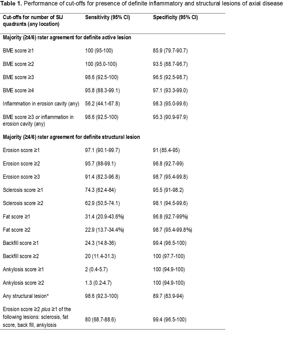
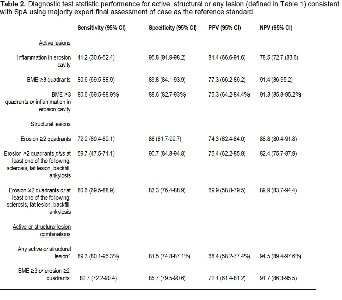
Results: Imaging from 243 subjects, 61% male, median age 14.9 had sequences available for detailed MRI scoring. The lesion-based cut-offs achieving specificity ≥95% with the highest sensitivity for a definite active inflammatory lesion were: BME in ≥3 quadrants or ≥1 inflammation in an erosion cavity (Table 1); combining the two types of lesions did not add incremental value. For definite structural lesion, the lesion cut-offs achieving ≥95% specificity with the highest sensitivity were the following: erosion in ≥2 quadrants, sclerosis in ≥1 quadrant, fat lesion in ≥1 quadrant, and backfill in ≥1 quadrant (Table 1); combinations of the lesions did not add incremental value. PPV and NPV of the quantitative threshold definitions for active, structural, or any lesions typical of axial SpA using the majority expert final assessment as the reference standard are shown in Table 2. The PPV and specificities were highest for BME ≥3 quadrants, erosion in ≥2 quadrants or a combination of the 2.

Conclusion: We propose cut-offs for definite active and structural lesions on MRI typical of axial disease in JSpA that have high PPV and specificity for clinical assessment. These cut-offs have applicability for diagnosis, therapeutic management, and classification.

*Any structural lesion with the following score thresholds: Erosion in ≥2 quadrants, sclerosis in ≥1 quadrant, fat lesion in ≥1 quadrant, backfill in ≥1 quadrant, or ankylosis ≥1 quadrant. Erosion SIJ quadrants = upper ilium, lower ilium, upper sacrum, lower sacrum for left and right SIJ; # of quadrants = total # of quadrants with detectable BME across all slices of study. Bone Marrow edema: An ill-defined area of high bone marrow signal intensity on fluid-sensitive sequences within the subchondral bone of the ilium or sacrum compared to the signal intensity of the iliac crest, edges of the vertebrae, and triradiate cartilage and in comparison to physiological changes normally seen on MRI examinations of age- and sex-matched children, and visible in 2 planes [1]. Inflammation at the site of an erosion: Increased signal on STIR and/or T1-weighted fat suppressed (FS) post-gadolinium at the site of erosion [2]. Erosion: A defect in subchondral bone associated with full-thickness loss of the dark appearance of the subchondral cortex at its expected location, with loss of signal on a T1-weighted sequence compared with the normal bright appearance of adjacent bone marrow [2]. Sclerosis: A substantially wider than normal (of ≥ 5 mm in adolescents) area of low subarticular bone signal on T1-weighted and fluid sensitive images [1]. Fat lesion: Homogeneous increased signal intensity within the subchondral bone on T1-weighted non-FS image presenting with a distinct border of the lesion [2]. Backfill: Bright signal on a T1-weighted sequence in a typical location for an erosion or confluent erosions, with signal intensity greater than normal bone marrow, which meets the following requirements: a) associated with complete loss of the dark appearance of the subchondral cortex at its expected location. B) clearly demarcated from adjacent bone marrow by an irregular band of dark signal reflecting sclerosis at the border of the original erosion [2]. Ankylosis: Presence of signal equivalent to regional bone marrow continuously bridging a portion of the joint space between the iliac and sacral bones [1]. BME = bone marrow edema; SIJ = sacroiliac joint; CI = confidence interval.

Confidence level was reported for each case on a scale of highly confident the case is not axial disease (_3) to highly confident the case is axial disease (+3). Cases rated as high confidence by the experts in having axial disease ≥+2 were used as the reference standard. ^Bone marrow edema in ≥3 quadrants, inflammation in an erosion cavity, Erosion in ≥2 quadrants, sclerosis, fat lesion, backfill, or ankylosis. PPV = positive predictive value; NPV = negative predictive value; CI = confidence interval; BME = bone marrow edema.


Disclosures: P. Weiss, Lilly, 1, 2, Pfizer, 1, 2, Biogen, 2; T. Brandon, None; A. Aggarwal, None; R. Burgos-Vargas, None; R. Colbert, Eli Lilly and Company, 12, Provide Drug; G. Horneff, Novartis, 5, 6, Janssen, 5, 6, Roche, 5, Eli-Lilly, 6, Glaxo Smith and Kline, 6, Pfizer, 6, Sobi, 6; R. Joos, None; R. Laxer, None; K. Minden, Abbvie, 6, Novartis, 2, 6, Sanofi, 2, Pfizer, 2; A. Ravelli, Abbvie, 2, 6, BMS, 2, 6, Pfizer, 2, 6, Hoffmann-La Roche, 2, 6, Novartis, 2, 6, Centocor, 2, 6, Anjelini Holding, 2, 6, Reckitt Benckiser, 2, 6; N. Ruperto, Ablynx, 2, 6, Amgen, 2, 6, Astrazeneca-Medimmune, 2, 6, Aurinia, 2, 6, Bayer, 2, 6, Bristol Myers and Squibb, 2, 6, 12, The IRCCS IGG, where NR works as full-time public employee has received contributions, this has been reinvested for research activities of the hospital in a fully independent manner, without any commitment with third parties, Cambridge Healthcare Research (CHR, 2, 6, Celgene, 2, 6, Domain therapeutic, 2, 6, Eli-Lilly, 2, 6, 12, The IRCCS IGG, where NR works as full-time public employee has received contributions, this has been reinvested for research activities of the hospital in a fully independent manner, without any commitment with third parties, EMD Serono, 2, 6, Glaxo Smith and Kline, 2, 6, Idorsia, 2, 6, Janssen, 2, 6, Novartis, 2, 6, 12, The IRCCS IGG, where NR works as full-time public employee has received contributions, this has been reinvested for research activities of the hospital in a fully independent manner, without any commitment with third parties, Pfizer, 2, 6, 12, The IRCCS IGG, where NR works as full-time public employee has received contributions, this has been reinvested for research activities of the hospital in a fully independent manner, without any commitment with third parties, Sobi, 2, 6, 12, The IRCCS IGG, where NR works as full-time public employee has received contributions, this has been reinvested for research activities of the hospital in a fully independent manner, without any commitment with third parties, UCB, 2, 6, F Hoffmann-La Roche, 12, The IRCCS IGG, where NR works as full-time public employee has received contributions, this has been reinvested for research activities of the hospital in a fully independent manner, without any commitment with third parties; J. Smith, None; M. Stoll, None; S. Tse, None; F. Van den Bosch, AbbVie, 2, 5, 6, Janssen, 2, 5, 6, UCB, 2, 5, 6, Bristol-Myers Squibb, 2, 6, Celgene, 2, 6, Eli Lilly, 2, 6, MSD, 2, 6, Novartis, 2, 6, Pfizer, 2, 6, Galapagos, 2, 6, Gilead, 2, 6; R. Lambert, Pfizer, 2; D. Biko, None; N. Chauvin, None; M. Francavilla, None; J. Jaremko, MEDO.ai, 8; N. Herregods, None; O. Kasapcopur, Novartis, 6, Pfizer, 6, Roche, 6, Abbvie, 6; M. YILDIZ, None; A. Hendry, None; W. Maksymowych, AbbVie, 2, 5, 6, Bristol-Myers Squibb, 2, 5, Boehringer Ingelheim, 2, Celgene, 2, 5, Eli Lilly, 2, 5, Galapagos, 2, 5, Janssen, 6, Novartis, 2, 5, 6, Pfizer Inc, 2, 5, 6, UCB, 2, 5, 6.

To cite this abstract in AMA style:

Weiss P, Brandon T, Aggarwal A, Burgos-Vargas R, Colbert R, Horneff G, Joos R, Laxer R, Minden K, Ravelli A, Ruperto N, Smith J, Stoll M, Tse S, Van den Bosch F, Lambert R, Biko D, Chauvin N, Francavilla M, Jaremko J, Herregods N, Kasapcopur O, YILDIZ M, Hendry A, Maksymowych W. Data-driven MRI Definitions for Active and Structural Sacroiliac Joint Lesions in Juvenile Spondyloarthritis Typical of Axial Disease [abstract]. Arthritis Rheumatol. 2021; 73 (suppl 9). https://acrabstracts.org/abstract/data-driven-mri-definitions-for-active-and-structural-sacroiliac-joint-lesions-in-juvenile-spondyloarthritis-typical-of-axial-disease/. Accessed .
  • Tweet
  • Click to email a link to a friend (Opens in new window) Email
  • Click to print (Opens in new window) Print

« Back to ACR Convergence 2021

ACR Meeting Abstracts - https://acrabstracts.org/abstract/data-driven-mri-definitions-for-active-and-structural-sacroiliac-joint-lesions-in-juvenile-spondyloarthritis-typical-of-axial-disease/

Advanced Search

Your Favorites

You can save and print a list of your favorite abstracts during your browser session by clicking the “Favorite” button at the bottom of any abstract. View your favorites »

All abstracts accepted to ACR Convergence are under media embargo once the ACR has notified presenters of their abstract’s acceptance. They may be presented at other meetings or published as manuscripts after this time but should not be discussed in non-scholarly venues or outlets. The following embargo policies are strictly enforced by the ACR.

Accepted abstracts are made available to the public online in advance of the meeting and are published in a special online supplement of our scientific journal, Arthritis & Rheumatology. Information contained in those abstracts may not be released until the abstracts appear online. In an exception to the media embargo, academic institutions, private organizations, and companies with products whose value may be influenced by information contained in an abstract may issue a press release to coincide with the availability of an ACR abstract on the ACR website. However, the ACR continues to require that information that goes beyond that contained in the abstract (e.g., discussion of the abstract done as part of editorial news coverage) is under media embargo until 10:00 AM ET on November 14, 2024. Journalists with access to embargoed information cannot release articles or editorial news coverage before this time. Editorial news coverage is considered original articles/videos developed by employed journalists to report facts, commentary, and subject matter expert quotes in a narrative form using a variety of sources (e.g., research, announcements, press releases, events, etc.).

Violation of this policy may result in the abstract being withdrawn from the meeting and other measures deemed appropriate. Authors are responsible for notifying colleagues, institutions, communications firms, and all other stakeholders related to the development or promotion of the abstract about this policy. If you have questions about the ACR abstract embargo policy, please contact ACR abstracts staff at [email protected].

Wiley

  • Online Journal
  • Privacy Policy
  • Permissions Policies
  • Cookie Preferences

© Copyright 2025 American College of Rheumatology