ACR Meeting Abstracts

ACR Meeting Abstracts

  • Meetings
    • ACR Convergence 2024
    • ACR Convergence 2023
    • 2023 ACR/ARP PRSYM
    • ACR Convergence 2022
    • ACR Convergence 2021
    • ACR Convergence 2020
    • 2020 ACR/ARP PRSYM
    • 2019 ACR/ARP Annual Meeting
    • 2018-2009 Meetings
    • Download Abstracts
  • Keyword Index
  • Advanced Search
  • Your Favorites
    • Favorites
    • Login
    • View and print all favorites
    • Clear all your favorites
  • ACR Meetings

Abstract Number: 849

Constructing a Macrophage Infiltration Timeline in a Murine Model of Osteoarthritis

Cheng Zhou1,2, Vu Nguyen1,3, Nisha Sambamurthy1,2, Michael Dodge1,4 and Carla Scanzello1,2, 1Translational Musculoskeletal Research Center, CMC Veterans Affairs Medical Center, Philadelphia, PA, 2Division of Rheumatology, University of Pennsylvania Perelman School of Medicine, Philadelphia, PA, 3University of Pennsylvania Perelman School of Medicine, Philadelphia, PA, 4Department of Orthopaedic Surgery, University of Pennsylvania Perelman School of Medicine, Philadelphia, PA

Meeting: 2018 ACR/ARHP Annual Meeting

Keywords: flow cytometry, Macrophage, Mouse model, osteoarthritis and synovium

  • Tweet
  • Click to email a link to a friend (Opens in new window) Email
  • Click to print (Opens in new window) Print
Session Information

Date: Sunday, October 21, 2018

Title: 3S085 ACR Abstract: Osteoarthritis & Joint Biology–Basic Science (846–850)

Session Type: ACR Concurrent Abstract Session

Session Time: 2:30PM-4:00PM

Background/Purpose:

Macrophage infiltration in synovium (SM) and intraarticular fat pads (FP) is common in Osteoarthritis (OA), and can contribute to catabolic cytokine and protease production as well as symptoms.  However, whether macrophages are targets for therapy in OA is unclear, as macrophages can also promote tissue repair. The purpose of this study is to characterize the timeline and subtypes of macrophages in SM and FP in a commonly used murine model of post-traumatic OA. 

Methods:

C57BL/6 male mice (10-12 wks old) were subjected to destabilization of medial meniscus (DMM) on the right leg.  For gene expression analysis, groups of mice were sacrificed pre-DMM (baseline), and post-DMM at 1, 2, 4, 8 and 16 weeks. Anterior SM and FP (combined) was dissected.  Tissues were pooled from 4-5 mice per sample to obtain adequate mRNA, cDNA synthesized by routine methods, and CD68 (a pan-macrophage marker) mRNA transcript number quantified using the QX200TM Droplet Digital PCR System (BioRad). Additional mice were sacrificed 4 and 8 weeks post-surgery and SM/FP dissected for cellular analysis.  Tissues from 4 knees were pooled, cells isolated enzymatically, and stained with the Live/Dead™ Fixable Violet Dead Cell Stain Kit (Invitrogen) and the following antibodies: CD45-PerCP Cy5.5, CD11c-Super Bright 645, F4/80-APC, iNOS-Alexa Fluor 488, CD206-PE. Multicolor flow cytometry was performed and data analyzed with FlowJo software (Version 10). Gating strategy is outlined in Fig 1A, and cells were expressed as percent of the CD45+ population.

Results:

CD68 mRNA level increased in DMM-operated limbs compared to preoperative levels, peaking at 4 weeks (21.61±3.02, Baseline: 2.24±0.42).   Levels remained significantly elevated compared to the un-operated limb up to 16 weeks (DMM: 4.77±0.31, contralateral: 0.90±0.26).  Percentages of F4/80+ macrophages and CD11c+ cells (expressed by murine monocytes and dendritic cells) in SM/FP were increased at 4 weeks post- DMM but not at 8 weeks (Fig 1B & C).  iNOS+ cells (primarily expressed on M1 macrophages) were slightly elevated compared to the un-operated side only at 8 weeks (p=0.02). Percentage of CD206+ cells (expressed on M2 macrophages) were similar in DMM and un-operated knees at either 4 or 8 weeks (Fig 1C). 

Conclusion:

Changes in macrophage populations can be detected in SM/FP tissues after DMM surgery by both gene expression and flow cytometry. CD68 gene expression was most pronounced early at 4 weeks, and paralleled changes in F4/80+ and CD11c+ cells. Flow cytometry of M1 and M2 markers suggested a slight increase in iNOS+ (M1-type) cells at 8 weeks and CD206 at 4 weeks.  Additional work is being pursued to further characterize macrophage phenotypes, and confirm gene expression seen at the chronic stage (16 weeks).  These observations will be used to plan timing of interventions to better understand the pathologic roles of macrophage subtypes in OA.

Figure 1


Disclosure: C. Zhou, None; V. Nguyen, None; N. Sambamurthy, None; M. Dodge, None; C. Scanzello, None.

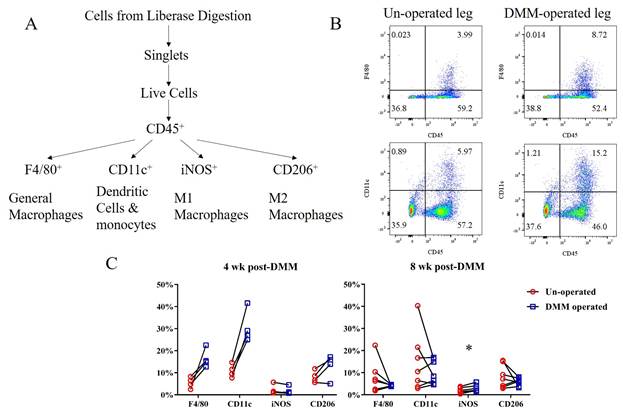
To cite this abstract in AMA style:

Zhou C, Nguyen V, Sambamurthy N, Dodge M, Scanzello C. Constructing a Macrophage Infiltration Timeline in a Murine Model of Osteoarthritis [abstract]. Arthritis Rheumatol. 2018; 70 (suppl 9). https://acrabstracts.org/abstract/constructing-a-macrophage-infiltration-timeline-in-a-murine-model-of-osteoarthritis/. Accessed .
  • Tweet
  • Click to email a link to a friend (Opens in new window) Email
  • Click to print (Opens in new window) Print

« Back to 2018 ACR/ARHP Annual Meeting

ACR Meeting Abstracts - https://acrabstracts.org/abstract/constructing-a-macrophage-infiltration-timeline-in-a-murine-model-of-osteoarthritis/

Advanced Search

Your Favorites

You can save and print a list of your favorite abstracts during your browser session by clicking the “Favorite” button at the bottom of any abstract. View your favorites »

All abstracts accepted to ACR Convergence are under media embargo once the ACR has notified presenters of their abstract’s acceptance. They may be presented at other meetings or published as manuscripts after this time but should not be discussed in non-scholarly venues or outlets. The following embargo policies are strictly enforced by the ACR.

Accepted abstracts are made available to the public online in advance of the meeting and are published in a special online supplement of our scientific journal, Arthritis & Rheumatology. Information contained in those abstracts may not be released until the abstracts appear online. In an exception to the media embargo, academic institutions, private organizations, and companies with products whose value may be influenced by information contained in an abstract may issue a press release to coincide with the availability of an ACR abstract on the ACR website. However, the ACR continues to require that information that goes beyond that contained in the abstract (e.g., discussion of the abstract done as part of editorial news coverage) is under media embargo until 10:00 AM ET on November 14, 2024. Journalists with access to embargoed information cannot release articles or editorial news coverage before this time. Editorial news coverage is considered original articles/videos developed by employed journalists to report facts, commentary, and subject matter expert quotes in a narrative form using a variety of sources (e.g., research, announcements, press releases, events, etc.).

Violation of this policy may result in the abstract being withdrawn from the meeting and other measures deemed appropriate. Authors are responsible for notifying colleagues, institutions, communications firms, and all other stakeholders related to the development or promotion of the abstract about this policy. If you have questions about the ACR abstract embargo policy, please contact ACR abstracts staff at [email protected].

Wiley

  • Online Journal
  • Privacy Policy
  • Permissions Policies
  • Cookie Preferences

© Copyright 2025 American College of Rheumatology