ACR Meeting Abstracts

ACR Meeting Abstracts

  • Meetings
    • ACR Convergence 2024
    • ACR Convergence 2023
    • 2023 ACR/ARP PRSYM
    • ACR Convergence 2022
    • ACR Convergence 2021
    • ACR Convergence 2020
    • 2020 ACR/ARP PRSYM
    • 2019 ACR/ARP Annual Meeting
    • 2018-2009 Meetings
    • Download Abstracts
  • Keyword Index
  • Advanced Search
  • Your Favorites
    • Favorites
    • Login
    • View and print all favorites
    • Clear all your favorites
  • ACR Meetings

Abstract Number: 1180

Confirmation of Manual Cartilage Segmentation Findings by Automated Segmentation: Retrospective Analysis of MRI Images from a Sprifermin Phase II Study

Alan Brett1, Michael A Bowes 1, Philip G Conaghan 2, Christoph Ladel 3, Jeffrey Kraines 4, Hans Guehring 3, Flavie Moreau 4 and Felix Eckstein 5, 1Imorphics, Manchester, United Kingdom, 2Leeds Institute of Rheumatic and Musculoskeletal Medicine, University of Leeds & NIHR Leeds Biomedical Research Centre, Leeds, United Kingdom, 3Merck KGaA, Darmstadt, Germany, 4EMD Serono Research and Development Institute, Inc. (a business of Merck KGaA, Darmstadt, Germany), Billerica, MA, 5Paracelsus Medical University, Salzburg, Austria

Meeting: 2019 ACR/ARP Annual Meeting

Keywords: cartilage, DMOAD, imaging techniques and osteoarthritis, Knee

  • Tweet
  • Click to email a link to a friend (Opens in new window) Email
  • Click to print (Opens in new window) Print
Session Information

Date: Monday, November 11, 2019

Title: Imaging Of Rheumatic Diseases Poster II

Session Type: Poster Session (Monday)

Session Time: 9:00AM-11:00AM

Background/Purpose: Sprifermin is under investigation as a potential disease-modifying osteoarthritis drug (DMOAD). 2-yr results from the FORWARD study showed significant dose-dependent modification of cartilage thickness in the total femorotibial joint (TFTJ), medial and lateral femorotibial joints (MFTC, LFTC), and central medial and lateral TFTJ subregions, by quantitative (q)MRI (Hochberg et al. ACR 2017).

This post hoc analysis aimed to determine whether qMRI findings from FORWARD (manual segmentation) could be reproduced in the same cartilage regions using an independent method (automated segmentation), on the same dataset/time period.

Methods: Pts were randomized 1:1:1:1:1 to: sprifermin 100 µg q6mo; 100 µg q12mo; 30 µg q6mo; 30 µg q12mo; and placebo (n=110/110/111/110/108). Cartilage thickness was assessed at baseline and 6, 12, 18, and 24 months using 1.5- or 3-Tesla MRI images, analysed manually. The same images were analysed by automated cartilage segmentation using active appearance models, a supervised machine learning method, to produce maps of cartilage thickness for weight-bearing femoral and tibial cartilage surfaces, subdivided into anatomical masks. Results were blinded for treatment and timepoint for both methods. No statistical comparisons between methods were conducted.

Endpoints were change from baseline in: 1) cartilage thickness in the TFTJ, MFTC and LFTC, using regions duplicated based on published data; 2) cartilage thickness in the central subregion of the medial and lateral tibia and femur (cMT, cMF, cLT, cLF [conventions used by the automated analysis investigators]). As in previous analyses, treatment effect was assessed by observed changes and adjusted using repeated ANCOVA on change from baseline, including treatment group, timepoint, and country as fixed factors, baseline value as covariate and treatment by timepoint as interaction.

Results: Based on automated segmentation, statistically significant, dose-dependent structural modification of cartilage thickness was observed over 2 yrs with sprifermin vs placebo for the TFTJ (overall treatment effect and dose response across all doses, both P< 0.001), MFTC (P=0.004 and P=0.044), and LFTC (both P< 0.001). Table 1 shows changes from baseline for sprifermin 100 µg q6mo and placebo. Statistically significant dose-dependent structural modification of cartilage over 2 yrs was observed for sprifermin vs placebo in the cMT (100 µg q6mo), cLT (100 µg q6mo, q12mo) and cLF (100 µg q6mo, q12mo). In the cMF, there was no treatment effect, but there was a linear trend for dose responsiveness. The results showed a consistent pattern to those obtained using manual segmentation.

Conclusion: Cartilage thickness assessed by automated segmentation provided a consistent pattern of structural modification in FORWARD compared with manual segmentation. This is the first time that two independent methods of image analysis have reached the same conclusions in an interventional DMOAD trial. The findings strengthen the conclusions that sprifermin modifies cartilage loss/structural progression in knee OA.
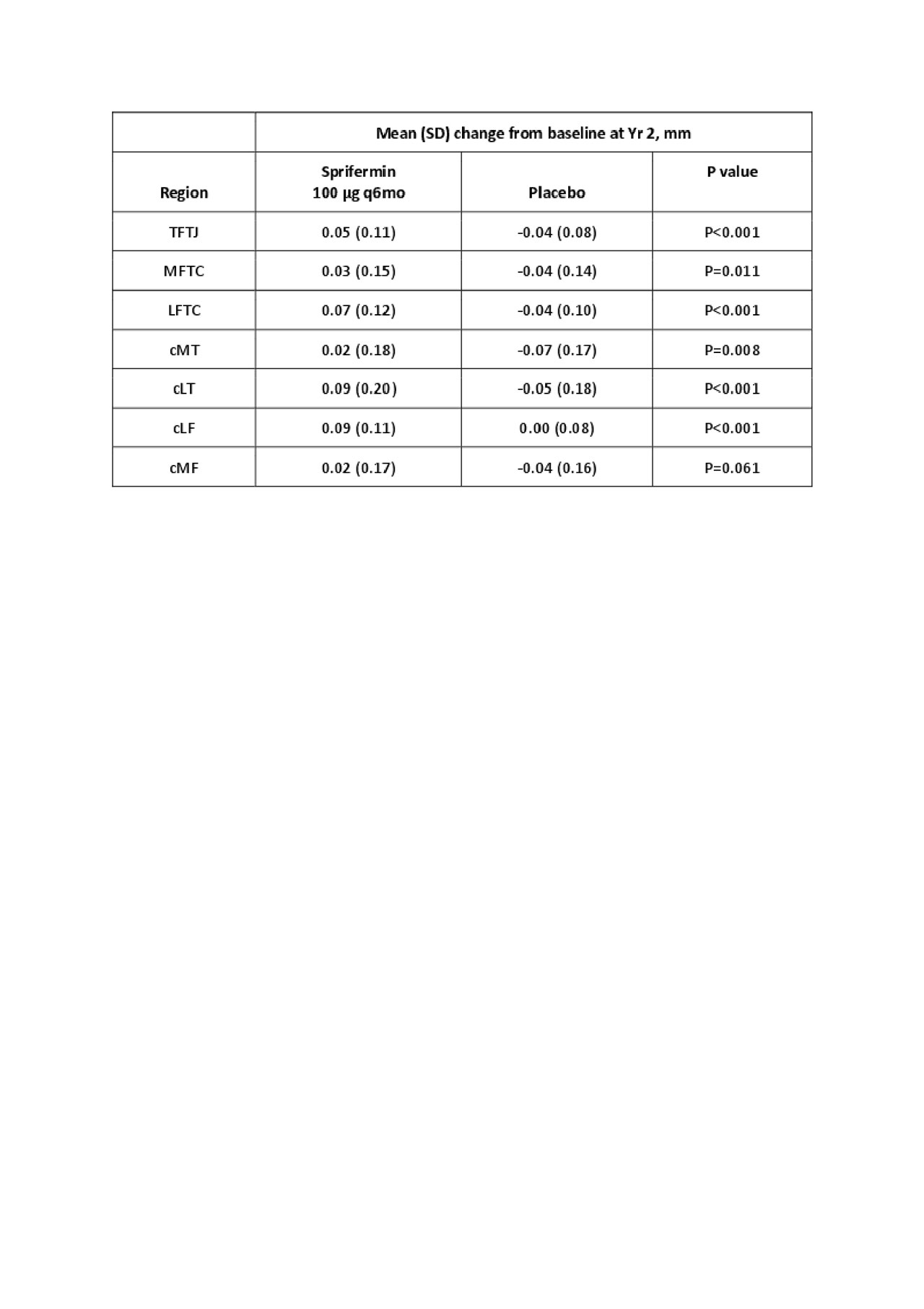

Table 1

Table 1. Change in cartilage thickness with sprifermin 100 µg q6mo vs placebo using automated segmentation


Disclosure: A. Brett, Imorphics, Manchester, UK, 3; M. Bowes, Imorphics, Manchester, UK, 3; P. Conaghan, Abbvie, 5, 8, AbbVie, 5, 8, AstraZeneca, 5, 8, BMS, 5, 8, Bristol Myers Squibb, 5, 8, Eli Lilly, 8, EMD, 5, EMD Serono, 5, EMD Serono Research and Development Institute, Inc., 5, 8, Flexion, 5, 8, Flexion Therapeutics, 5, 8, Galapagos, 5, 8, Glaxo Smith Kline, 5, GlaxoSmithKline, 5, 8, Lilly, 8, Medivir, 5, 8, Novartis, 5, 8, Pfizer, 5, 8, Roche, 5, 8, Samumed, 5, 8, Serono, 5, Stryker, 5, 8; C. Ladel, Merck KGaA, Darmstadt, Germany, 3; J. Kraines, EMD Serono Research and Development Institute, Inc., 3, EMD Serono Research and Development Institute, Inc. (a business of Merck KGaA, Darmstadt, Germany), 3; H. Guehring, Merck KGaA, Darmstadt, Germany, 3; F. Moreau, EMD Serono Research and Development Institute, Inc., 3, EMD Serono Research and Development Institute, Inc. (a business of Merck KGaA, Darmstadt, Germany), 3; F. Eckstein, Chrondrometrics GmbH, 1, 3, Merck KGaA, Darmstadt, Germany, 5, Samumed LLC, 5, Galapagos, 5, Abbvie, 5, Bioclinica, 5, Kolon TissueGene, 5, Novartis, 5, Servier, 5, Roche, 5.

To cite this abstract in AMA style:

Brett A, Bowes M, Conaghan P, Ladel C, Kraines J, Guehring H, Moreau F, Eckstein F. Confirmation of Manual Cartilage Segmentation Findings by Automated Segmentation: Retrospective Analysis of MRI Images from a Sprifermin Phase II Study [abstract]. Arthritis Rheumatol. 2019; 71 (suppl 10). https://acrabstracts.org/abstract/confirmation-of-manual-cartilage-segmentation-findings-by-automated-segmentation-retrospective-analysis-of-mri-images-from-a-sprifermin-phase-ii-study/. Accessed .
  • Tweet
  • Click to email a link to a friend (Opens in new window) Email
  • Click to print (Opens in new window) Print

« Back to 2019 ACR/ARP Annual Meeting

ACR Meeting Abstracts - https://acrabstracts.org/abstract/confirmation-of-manual-cartilage-segmentation-findings-by-automated-segmentation-retrospective-analysis-of-mri-images-from-a-sprifermin-phase-ii-study/

Advanced Search

Your Favorites

You can save and print a list of your favorite abstracts during your browser session by clicking the “Favorite” button at the bottom of any abstract. View your favorites »

All abstracts accepted to ACR Convergence are under media embargo once the ACR has notified presenters of their abstract’s acceptance. They may be presented at other meetings or published as manuscripts after this time but should not be discussed in non-scholarly venues or outlets. The following embargo policies are strictly enforced by the ACR.

Accepted abstracts are made available to the public online in advance of the meeting and are published in a special online supplement of our scientific journal, Arthritis & Rheumatology. Information contained in those abstracts may not be released until the abstracts appear online. In an exception to the media embargo, academic institutions, private organizations, and companies with products whose value may be influenced by information contained in an abstract may issue a press release to coincide with the availability of an ACR abstract on the ACR website. However, the ACR continues to require that information that goes beyond that contained in the abstract (e.g., discussion of the abstract done as part of editorial news coverage) is under media embargo until 10:00 AM ET on November 14, 2024. Journalists with access to embargoed information cannot release articles or editorial news coverage before this time. Editorial news coverage is considered original articles/videos developed by employed journalists to report facts, commentary, and subject matter expert quotes in a narrative form using a variety of sources (e.g., research, announcements, press releases, events, etc.).

Violation of this policy may result in the abstract being withdrawn from the meeting and other measures deemed appropriate. Authors are responsible for notifying colleagues, institutions, communications firms, and all other stakeholders related to the development or promotion of the abstract about this policy. If you have questions about the ACR abstract embargo policy, please contact ACR abstracts staff at [email protected].

Wiley

  • Online Journal
  • Privacy Policy
  • Permissions Policies
  • Cookie Preferences

© Copyright 2025 American College of Rheumatology