
Session Information
Date: Sunday, November 5, 2017
Title: Systemic Lupus Erythematosus – Clinical Aspects and Treatment Poster I: Biomarkers and Outcomes
Session Type: ACR Poster Session A
Session Time: 9:00AM-11:00AM
Background/Purpose: Abnormal activation of the complement system is emerging as a useful biomarker in the evaluation of patients presenting symptoms of autoimmune rheumatic diseases. We sought to evaluate the relationships between complement activation and autoantibodies associated with systemic lupus erythematosus (SLE), anti-phospholipid Syndrome (APS) and rheumatoid arthritis (RA).
Methods: From May 2014 to November 2016, a cohort of 32,404 patients within the United States (mean 51±15 [SD] years, 86% females) was tested. EDTA whole blood and serum were collected within 48 hours of patient examination and processed in a CLIA certified/CAP accredited clinical laboratory. C4d bound to erythrocyte or B lymphocyte above the 99th percentile of normal healthy group were defined as abnormal. The panel of 16 autoantibodies (all determined by solid phase immunoassays) consisted of 6 SLE autoantibodies (high ANA, anti-dsDNA confirmed using Crithidia, anti-U1 RNP, anti-C1q, anti-ribosomal P, anti-Smith, all IgGs), 6 APS autoantibodies (anti-cardiolipin, anti-β2 glycoprotein 1, anti-phosphatidylserine/prothrombin complex, IgM and IgG) and 4 RA autoantibodies (anti-CCP, anti-MCV, IgM RF, IgA RF). The relationships between abnormal complement activation and the presence of the autoantibodies were analyzed on de-identified patient data using multivariate logistic regression with abnormal complement activation as the dependent variable and the presence of autoantibodies as predictors. Adjusted odds ratio were calculated for each autoantibody.
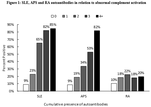
Results: Of the 32,404 patients tested 12% of them presented with abnormal complement activation. The overall incidence of autoantibodies ranged from 1% (anti-Sm) to 22% (ANA). The presence of SLE and APS antibodies were all associated with abnormal complement activation with adjusted OR ranging from 1.40 for anti-C1q (CI95%: 1.24-1.58) to 4.52 for anti-dsDNA (CI95%: 3.95-5.17), and from 1.47 for anti-cardiolipin IgG (CI95%: 1.22-1.77) to 3.2 for anti-PS/PT IgM (CI95%: 3.20-2.92), respectively (p<0.02). Of the 4 RA-associated antibodies, only anti-CCP (adjusted OR=1.25, CI95%: 1.02-1.54) and IgM RF (adjusted OR=1.17, CI95%: 1.04-1.32) were significantly associated with complement activation (p<0.05). Figure 1 illustrates the relationship between the cumulative presence of lupus, APS and RA autoantibodies, and abnormal complement activation. Across the cumulative range of SLE and APS associated antibodies, we detected a 319-fold (CI95%: 240-424), and 120-fold (CI95%: 94-153) increased likelihood of abnormal complement. In contrast, the cumulative presence of RA autoantibodies yielded minimum impact of the likelihood of abnormal complement activation (adjusted OR=2.3; CI95%: 2.0-2.7)
Conclusion: These diagnostic immunology data suggest that complement activation in peripheral blood is intimately related to SLE and APS antibodies.
Disclosure: T. Dervieux, Exagen, 3; J. Conklin, Exagen, 3; J. Ligayon, Exagen Diagnostics, Inc., 3; R. Lafon, Exagen Diagnostics, Inc., 3; A. Sace, Exagen Diagnostics, Inc., 3; T. O'Malley, Exagen Diagnostics, 3; R. Alexander, Exagen, 3; C. Ibarra, Exagen Diagnostics, Inc., 3.
To cite this abstract in AMA style:
Dervieux T, Conklin J, Ligayon J, Lafon R, Sace A, O'Malley T, Alexander R, Ibarra C. Complement Activation in Peripheral Blood in Relation to Lupus, Antiphospholipid and Rheumatoid Arthritis Autoantibodies: Insights from Clinical Laboratory Evaluations [abstract]. Arthritis Rheumatol. 2017; 69 (suppl 10). https://acrabstracts.org/abstract/complement-activation-in-peripheral-blood-in-relation-to-lupus-antiphospholipid-and-rheumatoid-arthritis-autoantibodies-insights-from-clinical-laboratory-evaluations/. Accessed .« Back to 2017 ACR/ARHP Annual Meeting
ACR Meeting Abstracts - https://acrabstracts.org/abstract/complement-activation-in-peripheral-blood-in-relation-to-lupus-antiphospholipid-and-rheumatoid-arthritis-autoantibodies-insights-from-clinical-laboratory-evaluations/