ACR Meeting Abstracts

ACR Meeting Abstracts

  • Home
  • Meetings Archive
    • ACR Convergence 2020
    • 2020 ACR/ARP PRSYM
    • 2019 ACR/ARP Annual Meeting
    • 2018 ACR/ARHP Annual Meeting
    • 2017 ACR/ARHP Annual Meeting
    • 2017 ACR/ARHP PRSYM
    • 2016-2009 Meetings
    • Download Abstracts
  • Keyword Index
  • Advanced Search
  • Your Favorites
    • Favorites
    • Login
    • Register
    • View and print all favorites
    • Clear all your favorites
  • Meeting Resource Center

Abstract Number: 2920

Comparison of Arterial Patterns of Disease in Takayasu’s Arteritis and Giant Cell Arteritis

K Bates Gribbons1, Cristina Ponte 2, Anthea Craven 3, David Cuthbertson 4, Simon Carette 5, Gary S. Hoffman 6, Nader A. Khalidi 7, Curry L. Koening 8, Carol Langford 9, Kathleen Maksimowicz-McKinnon 10, Carol A. McAlear 11, Paul Monach 12, Larry Moreland 13, Christian Pagnoux 14, Kaitlin Quinn 15, Joanna Robson 16, Philip Seo 17, Antoine Sreih 18, Ravi Suppiah 19, Kenneth Warrington 20, Steven Ytterberg 21, Raashid Luqmani 3, Richard Watts 22, Peter Merkel 18 and Peter C. Grayson 23, 1National Institute of Arthritis and Musculoskeletal and Skin Diseases, National Institutes of Health, Bethesda, MD, 2Department of Rheumatology, Hospital de Santa Maria, Lisbon, Portugal, 3University of Oxford, Oxford, United Kingdom, 4University of South Florida, Tampa, FL, 5Division of Rheumatology, Mount Sinai Hospital, University of Toronto, Toronto, Canada, 6Cleveland Clinic Foundation, Cleveland, OH, 7McMaster University, Hamilton, ON, Canada, 8University of Utah Hospital, Salt Lake City, UT, 9Cleveland Clinic, Cleveland, OH, 10Henry Ford Hospital, Wayne State University, Detroit, MI, 11University of Pennsylvania - VCRC Project Manager, Philadelphia, PA, 12Brigham and Women's Hospital, Boston, MA, 13University of Pittsburgh, PITTSBURGH, PA, 14Mount Sinai Hospital and University Health Network, Toronto, ON, Canada, 15Georgetown University Hospital/National Institutes of Health, Washington, DC, 16Faculty of Health and Applied Sciences, University of the West of England, Bristol, United Kingdom, 17Johns Hopkins Medicine, Baltimore, MD, 18University of Pennsylvania, Philadelphia, PA, 19Department of Rheumatology, Auckland District Health Board, Auckland, New Zealand, 20Mayo Clinic Rochester, Rochester, MN, 21Mayo Clinic College of Medicine, Rochester, MN, 22Norwich Medical School, University of East Anglia, Norwich, United Kingdom, 23National Institute of Arthritis and Musculoskeletal and Skin Diseases (NIAMS) of the National Institutes of Health, Bethesda, MD, Bethesda, MD

Meeting: 2019 ACR/ARP Annual Meeting

Keywords: 18FDG PET/CT scan and Angiography, giant cell arteritis, large vessel vasculitis, Takayasu arteritis

  • Tweet
  • Email
  • Print
Save to PDF
Session Information

Date: Wednesday, November 13, 2019

Session Title: 6W024: Vasculitis – Non-ANCA-Associated & Related Disorders III: Large Vessel Vasculitis Pathogenesis & Imaging (2918–2923)

Session Type: ACR Abstract Session

Session Time: 11:00AM-12:30PM

Background/Purpose: Current classification criteria differentiate between Takayasu’s arteritis (TAK) and giant cell arteritis (GCA), the two most common forms of large-vessel vasculitis, based primarily on clinical assessment, yet patients with TAK and GCA may differ in patterns of arterial disease.  This study aimed to use computer-based algorithms to determine if patterns of arterial disease are useful to differentiate TAK from GCA with large-vessel involvement (LV-GCA).

Methods: Patients with TAK or LV-GCA were studied from four independent cohorts: a large, international cohort and three separate cohorts combined into one North America cohort.  Case inclusion required evidence of large-vessel involvement, defined as stenosis, occlusion, or aneurysm by imaging or catheter-based angiography or ultrasound, or increased FDG uptake by positron-emission tomography (PET) in at least one of 11 specified arterial territories.  K-means cluster analysis was performed to identify clusters of patients based on pattern of arterial involvement.  Cluster groups were identified in the international cohort and independently validated in the combined North American cohort.

Results: A total of 1,068 patients were included (International: TAK=461, GCA=217; NA: TAK=225, GCA=165).  Patients with TAK underwent angiography (95%), ultrasonography (28%), and/or PET imaging (14%).  Patients with LV-GCA underwent angiography (50%), ultrasonography (52%), and/or PET imaging (58%).  Six distinct clusters of patients were identified in the international cohort and validated in the North American cohort (Figure).  Patients in Clusters One, Two, and Three were significantly more likely to have TAK, and patients in Clusters Four, Five, and Six were significantly more likely to have LV-GCA.  Out of all study patients, involvement of the abdominal aorta and renal/mesenteric arteries was the most specific pattern for TAK (TAK: 134 (92%) vs GCA: 11 (8%), p< 0.01), while bilateral subclavian/axillary disease was the most specific pattern for GCA (GCA: 92 (80%) vs TAK: 23 (20%),  p< 0.01).  Among patients who underwent both angiography and PET, patients with TAK were more likely than patients with GCA to have damage by angiography without associated inflammation by PET (TAK: 52% vs GCA: 7%, p< 0.001), and patients with GCA were more likely than patients with TAK to have arterial FDG-uptake by PET without associated vascular damage (GCA: 80% vs TAK: 23%, p< 0.001).

Conclusion: These findings support the incorporation of arterial patterns of disease into classification criteria for large-vessel vasculitis and highlight shared and divergent vascular phenotypes between TAK and GCA.


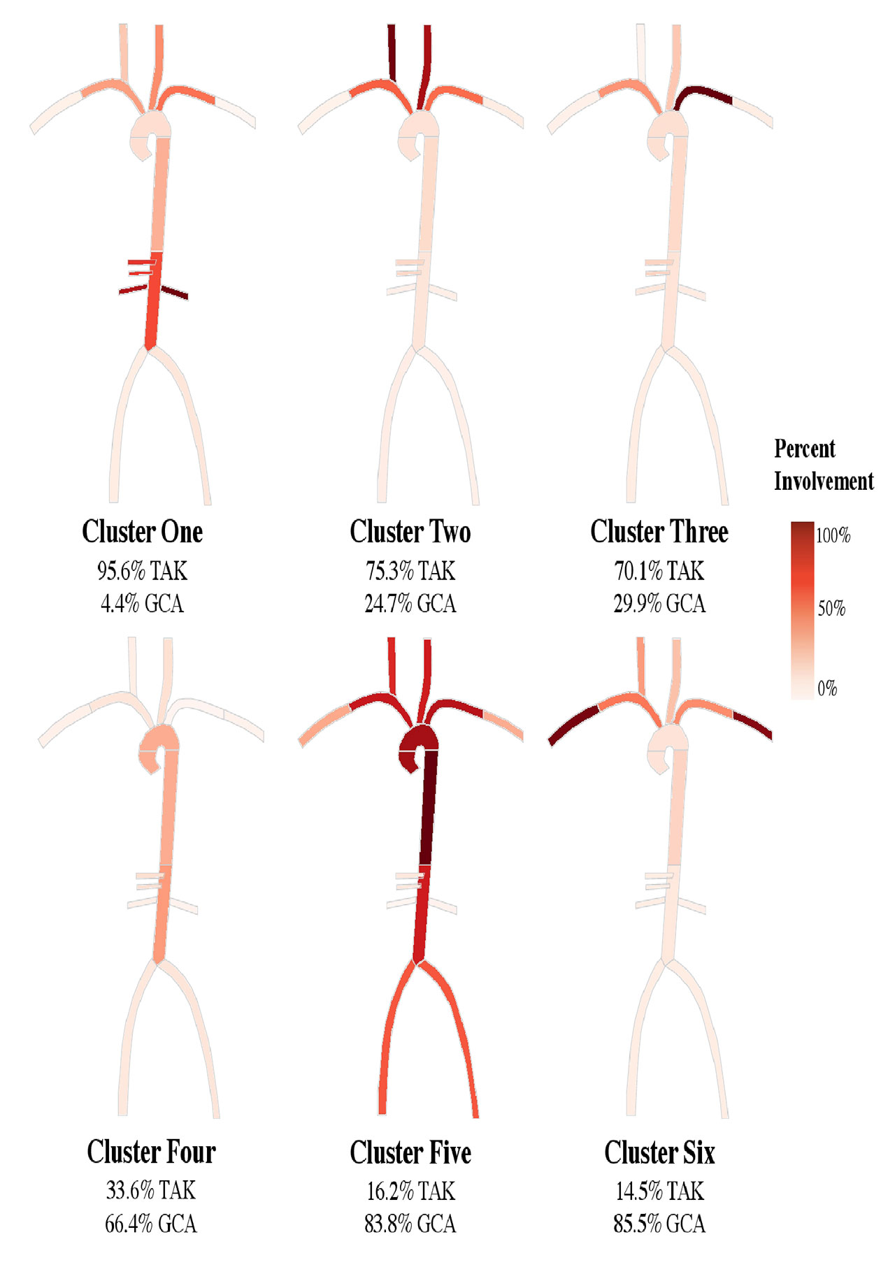
2019.03.14 TAK and GCA Imaging – Figures

Heatmaps of the large arteries indicating the percent of patients in each cluster from the international cohort with involvement of the 11 arterial territories of interest -right and left carotid, subclavian, axillary, and renal arteries, mesenteric arteries, and descending and abdominal aorta-. Increasingly darker red indicates an increasing percentage of patients in the cluster have involvement of the artery. The weighted proportion of patients with Takayasu’s arteritis -TAK- and patients with giant cell arteritis -GCA- in each cluster are listed. Clusters One, Two, and Three were specific for TAK in the International -development- and North American -validation- cohorts. Clusters Four, Five, and Six were specific for GCA in the International cohort but only Clusters Four and Six was specific for GCA in the North American cohort.


Disclosure: K. Gribbons, None; C. Ponte, None; A. Craven, None; D. Cuthbertson, None; S. Carette, None; G. Hoffman, None; N. Khalidi, None; C. Koening, None; C. Langford, Bristol-Myers Squibb, 2, GlaxoSmithKline,, 2, ChemoCentryx, 2, Genentech, 2, Bristol-Myers Squibb, 5, 9, Abbvie, 9, AstraZeneca, 9; K. Maksimowicz-McKinnon, AstraZeneca, Gilead, GSK, Merck, 9, ChemoCentryx, 5; C. McAlear, None; P. Monach, None; L. Moreland, None; C. Pagnoux, ChemoCentryx, 5, Chemocentryx, 5, Genetech/Roche, 5, Genzyme/Sanofi, 5, GlaxoSmithKline, 5, Hoffman-La Roche, 2, 5, 8, Hoffman-LaRoche, 2, 5, 8, Sanofi, 5; K. Quinn, None; J. Robson, None; P. Seo, None; A. Sreih, Bristol-Meyers Squibb, 3, Bristol-Myers Squibb, 3; R. Suppiah, None; K. Warrington, Eli Lilly, 2, GlaxoSmithKline, 2, roche, 2, 8, Roche, 2, 5, 8, Sanofi, 5, sanofi, 5; S. Ytterberg, None; R. Luqmani, Roche, 8, Roche, Vifor, InflaRx, 2; R. Watts, None; P. Merkel, Abbvie, 5, AbbVie, 5, AstraZeneca, 2, 5, AstraZeneca,, 2, 5, Biogen, 5, Boeringher-Ingelheim, 2, 5, Bristol-Myers Squibb, 2, 5, Celegene, 2, 5, Celgene, 2, 5, ChemoCentryx, 2, 5, CSL Behring, 5, Genentech/Roche, 2, 5, Genetech/Roche, 2, 5, Genzyme/Sanofi, 2, 5, GlaxoSmithKline, 2, 5, InflaRx, 5, Insmed, 5, Jannsen, 5, Kiniksa, 5, Kypha, 2, TerumoBCT, 2, UpToDate, 7; P. Grayson, None.

To cite this abstract in AMA style:

Gribbons K, Ponte C, Craven A, Cuthbertson D, Carette S, Hoffman G, Khalidi N, Koening C, Langford C, Maksimowicz-McKinnon K, McAlear C, Monach P, Moreland L, Pagnoux C, Quinn K, Robson J, Seo P, Sreih A, Suppiah R, Warrington K, Ytterberg S, Luqmani R, Watts R, Merkel P, Grayson P. Comparison of Arterial Patterns of Disease in Takayasu’s Arteritis and Giant Cell Arteritis [abstract]. Arthritis Rheumatol. 2019; 71 (suppl 10). https://acrabstracts.org/abstract/comparison-of-arterial-patterns-of-disease-in-takayasus-arteritis-and-giant-cell-arteritis/. Accessed March 1, 2021.
  • Tweet
  • Email
  • Print
Save to PDF

« Back to 2019 ACR/ARP Annual Meeting

ACR Meeting Abstracts - https://acrabstracts.org/abstract/comparison-of-arterial-patterns-of-disease-in-takayasus-arteritis-and-giant-cell-arteritis/

Advanced Search

Your Favorites

You can save and print a list of your favorite abstracts by clicking the “Favorite” button at the bottom of any abstract. View your favorites »

ACR Convergence: Where Rheumatology Meets. All Virtual. November 5-9.

ACR Pediatric Rheumatology Symposium 2020

© COPYRIGHT 2021 AMERICAN COLLEGE OF RHEUMATOLOGY

Wiley

  • Home
  • Meetings Archive
  • Advanced Search
  • Meeting Resource Center
  • Online Journal
  • Privacy Policy
  • Permissions Policies
loading Cancel
Post was not sent - check your email addresses!
Email check failed, please try again
Sorry, your blog cannot share posts by email.
This site uses cookies: Find out more.