Session Information
Session Type: Abstract Submissions (ACR)
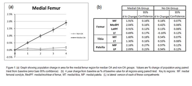
Background/Purpose: Change in subchondral bone has been clinically associated with progression of osteoarthritis (OA). Modern image analysis techniques allow accurate, automated identification of bone in MR images, facilitating the use of 3D changes in the bone to characterise and monitor OA. Objective was to compare rate of change in bone area of all the knee bones from all subjects in the OAI dataset with definite medial OA with a control group absent of radiological OA over a 4 year period.
Methods: 933 subjects with medial OA and MR images at baseline, 12, 24, 36 and 48 month were selected from the Osteoarthritis Initiative dataset; medial OA was defined as KL≥ 2 and presence of medial osteophytes. 904 control subjects with absence of radiographic OA were also identified, defined as KL=0 at all time-points. One index knee was analyzed per subject. Femur, tibia and patella bones were automatically segmented from MRIs using active appearance models1. Anatomical areas were automatically identified within the model2 and were measured at each time-point. All regions of the articulating surface of the femur, tibia and patella were included in the analysis.
Results: Mean age (SD) of the case group was 62 years (8.8); control group 59 (8.9); mean (SD) BMI for case/control 29.7(4.9)/26.9(4.3); %females for case/control 35%/47%. Rate of change of bone area in the medial compartments was typically 0.5% per annum in the case group with significantly lower change in the controls. Lateral compartments exhibited around half this amount of change, and showed less difference in the rates of change between cases and controls (though still highly significant). ANCOVA analysis demonstrated that age, BMI and gender could explain only a small amount of the variance in bone change.
Conclusion: Change in bone area within all knee compartments discriminated significantly between OA and control subjects. Discrimination was strongest in medial compartments, and particularly in the femur. The patella-femoral joint is an important component of OA disease, and discriminates between the groups as strongly as the femorotibial joint. Measurement of bone change provides a valuable tool for monitoring OA progression.
References:
1 Cootes, T. F, G. J. Edwards, and C. J. Taylor. “Active Appearance Models.” IEEE Trans Patt Anal Mach Intell 23.6 (2001): 681-85.
2 Williams, T. G., et al. Br.J.Radiol. 83.995 (2010): 940-48.
Disclosure:
M. A. Bowes,
Imorphics Ltd,
1,
Imorphics Ltd,
3;
C. B. Wolstenholme,
Imorphics Ltd,
3,
Imorphics Ltd,
1;
D. Hopkinson,
None;
G. R. Vincent,
Imorphics Ltd,
3,
Imorphics Ltd,
1;
P. G. Conaghan,
None.
« Back to 2012 ACR/ARHP Annual Meeting
ACR Meeting Abstracts - https://acrabstracts.org/abstract/changes-in-subchondral-bone-provide-a-sensitive-marker-for-osteoarthritis-and-its-progression-results-from-a-large-osteoarthritis-initiative-cohort/

