ACR Meeting Abstracts

ACR Meeting Abstracts

  • Meetings
    • ACR Convergence 2024
    • ACR Convergence 2023
    • 2023 ACR/ARP PRSYM
    • ACR Convergence 2022
    • ACR Convergence 2021
    • ACR Convergence 2020
    • 2020 ACR/ARP PRSYM
    • 2019 ACR/ARP Annual Meeting
    • 2018-2009 Meetings
    • Download Abstracts
  • Keyword Index
  • Advanced Search
  • Your Favorites
    • Favorites
    • Login
    • View and print all favorites
    • Clear all your favorites
  • ACR Meetings

Abstract Number: 65

CD6 Modulation Ameliorates Skin and Kidney Disease in a Spontaneous Murine Model of SLE

Samantha Chalmers1, Sayra Garcia 1, Rajalakshmy Ayilam Ramachandran 2, Chandra Mohan 2, Jeanette Ampudia 3, Cherie Ng 3, Stephen Connelly 3 and Chaim Putterman 4, 1Albert Einstein College of Medicine, Bronx, NY, 2University of Houston, Houston, 3Equillium, Inc, San Diego, CA, 4Albert Einstein College of Medicine, New York, NY

Meeting: 2019 ACR/ARP Annual Meeting

Keywords: cutaneous lupus, Lupus nephritis, SLE, T cells and Lupus

  • Tweet
  • Email
  • Print
Session Information

Date: Sunday, November 10, 2019

Title: SLE – Animal Models Poster

Session Type: Poster Session (Sunday)

Session Time: 9:00AM-11:00AM

Background/Purpose: T cells are an important contributor the pathogenesis of SLE and its various end organ manifestations. Thus, they present themselves as potential therapeutic targets; however, the optimal approach to targeting T cells in SLE still remains under investigation. CD6 is a co-stimulatory receptor, predominantly expressed on T cells, that binds to activated leukocyte cell adhesion molecule (ALCAM), a ligand expressed on antigen presentation cells and various epithelial and endothelial tissues.  The CD6-ALCAM pathway plays an integral role in modulating T cell activation, proliferation, differentiation and trafficking. In this study, we assessed the expression of CD6 and ALCAM within the context of a murine model of SLE, and then subsequently targeted this signaling axis to determine its role in the pathogenesis of disease.  

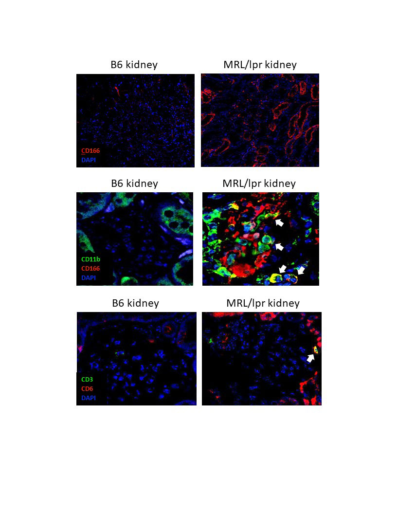
Methods: The MRL/lpr mouse is a spontaneous murine model of SLE, which exhibits systemic autoimmunity, nephritis, and skin disease. In an initial experiment, 6 month old MRL/lpr mice and B6 kidneys were stained for the presence of both ALCAM and CD6. In a separate experiment, female MRL/lpr mice were aged to 9-10 weeks of age, and then treated with either anti-CD6 monoclonal antibody (60 ug/dose, IP twice per week), isotype control (60 ug/dose, twice per week), or cyclophosphamide (25 mg/kg, once per week). We also included a no treatment group, and a group of MRL/MpJ mice, a congenic healthy control strain. Baseline levels of anti-DNA antibodies, weight, and proteinuria in the MRL/lpr groups were similar. Mice were monitored weekly for proteinuria, lymph node swelling, and macroscopic skin lesions.

Results: MRL/lpr mice at 6 months of age show increased levels of ALCAM expression in their kidneys, both within their tubules and glomeruli, compared to B6 healthy control mice (shown are images representative of 3 mice per group). Additionally, macrophages infiltrating into the glomeruli of MRL/lpr mice were ALCAM+ (white arrows, top panel) and were paired with a concomitant increase in CD6+ T cell infiltration (white arrows, bottom panel). Consequently, in a separate study, we blocked this signaling axis using a monocolonal antibody for CD6. At 19 weeks of age, mice treated with anti-CD6 show improved proteinuria compared to isotype control mice (p< 0.05). MRL/lpr mice develop lymphoproliferative disease which results in abnormally large lymph nodes. Assessing lymphadenopathy at 19 weeks of age, we noted a significant improvement in the anti-CD6 treated mice, compared to the isotype control (p< 0.05). MRL/lpr mice also develop severe skin lesions that have similar pathology to cutaneous lupus. Macroscopic scoring of these lesions showed a significant improvement in the skin disease of the anti-CD6 treated mice compared to the isotype control group (p< 0.05).

Conclusion: Within a spontaneous model of SLE, anti-CD6 treatment ameliorated multiple end organ pathologies, namely in the kidney and skin, while also significantly reducing the lymphoproliferative phenotype of this model. Overall, these results indicate that targeting the CD6-ALCAM pathway may have promising therapeutic potential for multiple end organ pathologies within SLE.

Upregulated expression of ALCAM -CD166, red-, is seen in the tubules of 6 month old MRL/lpr mice, compared to age matched B6 mice -top panel-. Looking more closely at the glomeruli, we note ALCAM expression is increased in lupus mice, including on macrophages -CD11b+, white arrows- infiltrating into the glomerulus -middle panel-. Finally, CD6+CD3+ T cells are also noted infiltrating the glomeruli of MRL/lpr mice -white arrow, bottom panel-. Shown are representative images from 3 mice per group.


Disclosure: S. Chalmers, Equillium, Inc, 2; S. Garcia, None; R. Ayilam Ramachandran, None; C. Mohan, Equillium, 5, Equillium, Inc, 5; J. Ampudia, Equillium, Inc, 3; C. Ng, Equillium, Inc, 3; S. Connelly, Equillium, Inc, 3, 6, Equillium, 3; C. Putterman, Equillium, 5, Equillium, Inc, 2, 5, Exagen, 2.

To cite this abstract in AMA style:

Chalmers S, Garcia S, Ayilam Ramachandran R, Mohan C, Ampudia J, Ng C, Connelly S, Putterman C. CD6 Modulation Ameliorates Skin and Kidney Disease in a Spontaneous Murine Model of SLE [abstract]. Arthritis Rheumatol. 2019; 71 (suppl 10). https://acrabstracts.org/abstract/cd6-modulation-ameliorates-skin-and-kidney-disease-in-a-spontaneous-murine-model-of-sle/. Accessed .
  • Tweet
  • Email
  • Print

« Back to 2019 ACR/ARP Annual Meeting

ACR Meeting Abstracts - https://acrabstracts.org/abstract/cd6-modulation-ameliorates-skin-and-kidney-disease-in-a-spontaneous-murine-model-of-sle/

Advanced Search

Your Favorites

You can save and print a list of your favorite abstracts during your browser session by clicking the “Favorite” button at the bottom of any abstract. View your favorites »

All abstracts accepted to ACR Convergence are under media embargo once the ACR has notified presenters of their abstract’s acceptance. They may be presented at other meetings or published as manuscripts after this time but should not be discussed in non-scholarly venues or outlets. The following embargo policies are strictly enforced by the ACR.

Accepted abstracts are made available to the public online in advance of the meeting and are published in a special online supplement of our scientific journal, Arthritis & Rheumatology. Information contained in those abstracts may not be released until the abstracts appear online. In an exception to the media embargo, academic institutions, private organizations, and companies with products whose value may be influenced by information contained in an abstract may issue a press release to coincide with the availability of an ACR abstract on the ACR website. However, the ACR continues to require that information that goes beyond that contained in the abstract (e.g., discussion of the abstract done as part of editorial news coverage) is under media embargo until 10:00 AM ET on November 14, 2024. Journalists with access to embargoed information cannot release articles or editorial news coverage before this time. Editorial news coverage is considered original articles/videos developed by employed journalists to report facts, commentary, and subject matter expert quotes in a narrative form using a variety of sources (e.g., research, announcements, press releases, events, etc.).

Violation of this policy may result in the abstract being withdrawn from the meeting and other measures deemed appropriate. Authors are responsible for notifying colleagues, institutions, communications firms, and all other stakeholders related to the development or promotion of the abstract about this policy. If you have questions about the ACR abstract embargo policy, please contact ACR abstracts staff at [email protected].

Wiley

  • Online Journal
  • Privacy Policy
  • Permissions Policies
  • Cookie Preferences

© Copyright 2025 American College of Rheumatology