Session Information
Session Type: Poster Session A
Session Time: 10:30AM-12:30PM
Background/Purpose: Osteoarthritis is associated with bone changes such as subchondral sclerosis, bone marrow lesions, and osteophyte formation (Donel S, 2019). Prior work has demonstrated that CD14-deficient mice show significantly less OA-associated subchondral bone (Sambamurthy N, 2018). CD14 is a GPI-anchored surface protein and co-receptor for several inflammatory TLRs, and is highly expressed in myeloid cells including osteoclast precursors (Zanoni, I 2013; Xue, J 2020). TLR activation can both activate and inhibit osteoclastogenic potential. Therefore, using a CD14-deficient mouse model we hypothesized that inhibitory effects of TLR-stimuli on osteoclast differentiation would be ameliorated in CD14 deficient cells.
Methods: Cell isolation and culture (n=3-5): Bone Marrow was isolated from femurs and tibiae of skeletally mature (10-12 wk old) C57BL/6 (WT) and CD14-deficient mice. Following 24 hr suspension culture, cells were cultured in complete aMEM with 30 ng/mL M-CSF, for 5 days to expand osteoclast precursors (macrophages). Cells were passaged on day 6 and cultured (24 well plate, 50,000 cells/well) in the presence or absence of RANKL (100 ng/mL). In a separate study, cells were stimulated with a TLR4-stimulus (LPS, 1 ng/mL), soluble CD14 (1 µg/mL), and a TLR4-inhibitor (CLI-095, 1 µg/mL), or an Anti-IFNAR1 antibody (1 ug/mL).
Osteoclast staining and image analysis: Cells were stained for Tartrate-resistant acid phosphatase (TRAP) on days 3 and 4 after addition of RANKL. Cells were imaged at 10x (3 images/well over 4 wells per timepoint) and quantified (%area covered) using ImageJ and CellProfiler. Number of osteoclasts (cells with >3 nuclei) per 10X field was also quantitated. Multiple unpaired t-test were performed with Holm-Sidak correction.
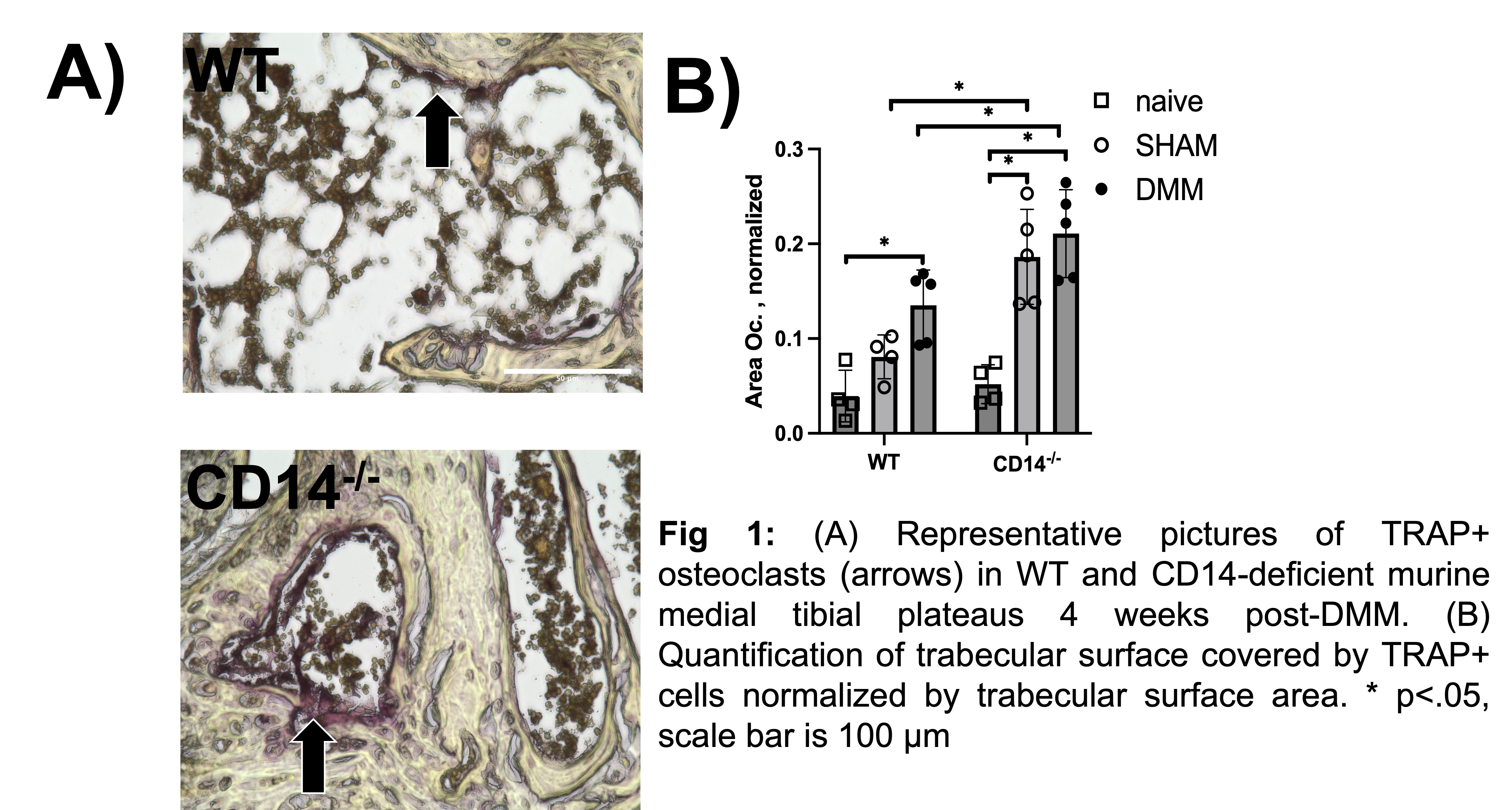
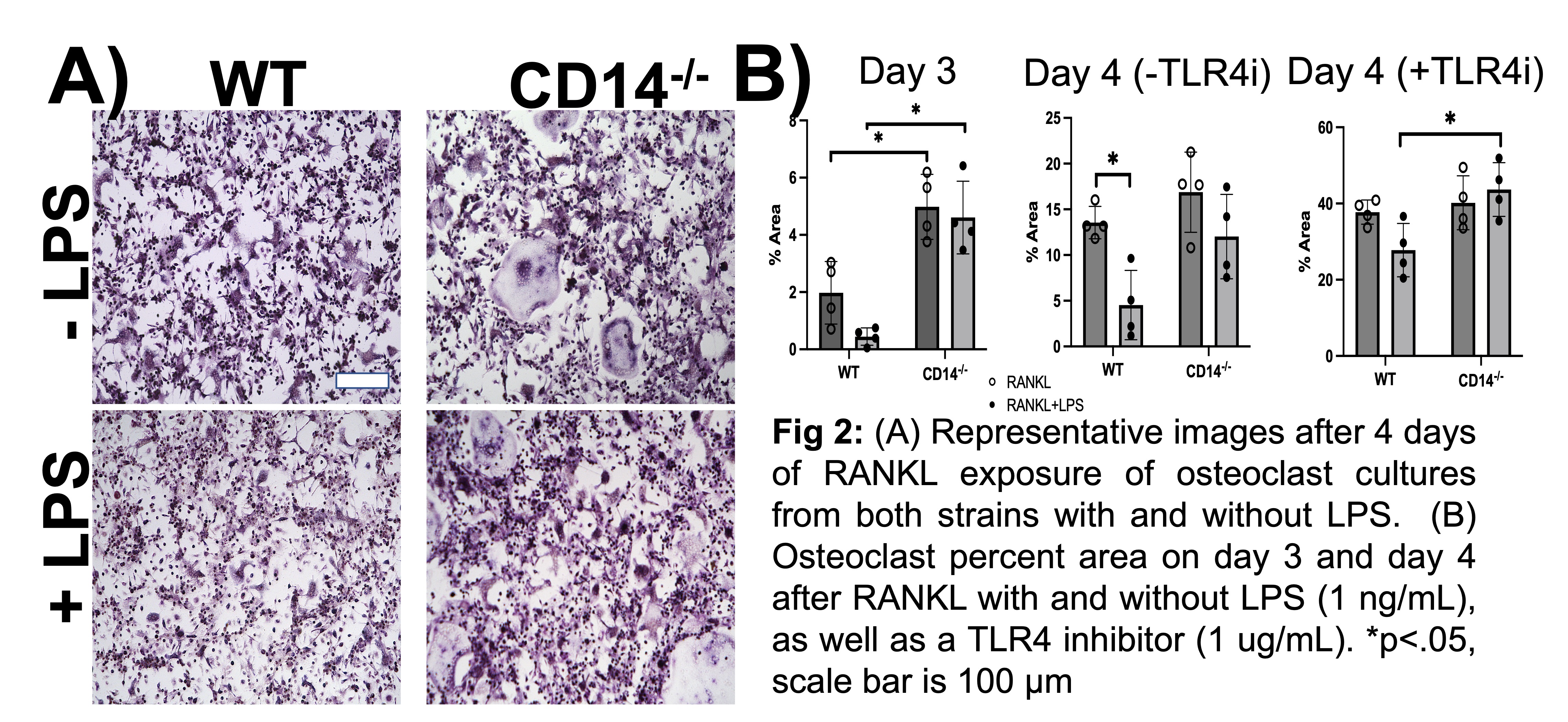
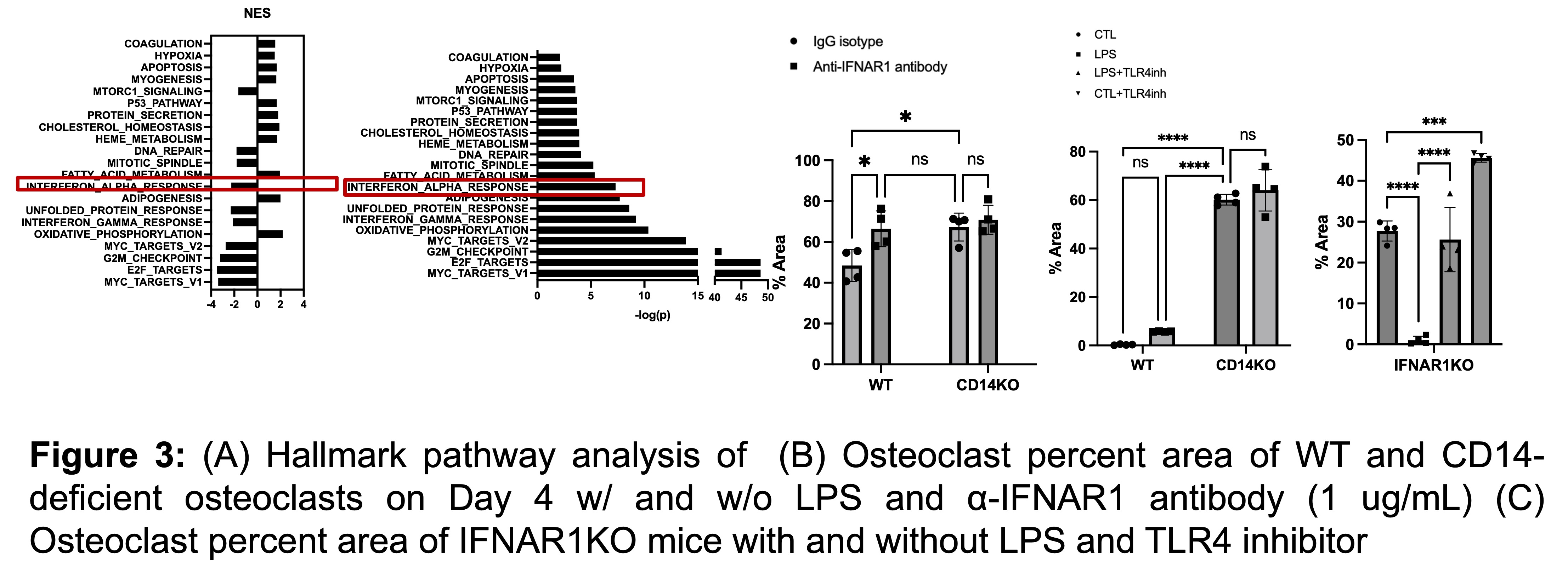
Results: CD14-deficient mice possessed increase osteoclast area at baseline and post-injury (Fig. 1A,B). Cells showed more rapid differentiation than WT cells at baseline (Fig. 2). Area of the plate covered by osteoclasts (Fig. 2B) were higher in CD14-deficient cells on day 3 and day 4. TLR4-stimuli: With LPS stimulation, CD14-deficient cells differentiated more quickly compared to WT at day 3 (p< 0.001) (Fig. 3G). LPS stimulation led to a 77% decrease of osteoclastogenesis in the WT cells, but only a 7.5% decrease in osteoclastogenesis on the CD14-deficient cells (Figure 2G, 3G) Addition of CLI-095 mitigated some inhibitory effects of LPS in WT, but had a synergistic effect on CD14-deficient cells (Fig 3H). The addition of sCD14 inhibited osteoclastogenesis in the CD14-deficient group, both with and without LPS. RNA sequencing was completed on osteoclasts on day 4 and showed decreased Type I Interferon signaling. At baseline, the Anti-IFNAR antibody mitigated the difference between WT and CD14-deficient mice. However, this was not shown with LPS addition.
Conclusion: Our results show that at baseline, CD14-deficient osteoclasts precursors differentiate more quickly than WT cells likely due to Type I Interferon singaling. Further, during TLR4 stimulation studies, CD14-deficient cells were protected from LPS-TLR4 mediated inhibition of osteoclastogenesis, compared to WT cells, likely due to decreased signaling through the MyD88-dependent pathway.
To cite this abstract in AMA style:
Murphy L, Burt K, Nguyen V, Hu B, Mauck R, Scanzello C. CD14 Deficiency Protects Against TLR4/LPS-mediated Inhibition of Osteoclastogenesis [abstract]. Arthritis Rheumatol. 2024; 76 (suppl 9). https://acrabstracts.org/abstract/cd14-deficiency-protects-against-tlr4-lps-mediated-inhibition-of-osteoclastogenesis/. Accessed .« Back to ACR Convergence 2024
ACR Meeting Abstracts - https://acrabstracts.org/abstract/cd14-deficiency-protects-against-tlr4-lps-mediated-inhibition-of-osteoclastogenesis/


