Session Information
Session Type: ACR Poster Session A
Session Time: 9:00AM-11:00AM
Background/Purpose: Wrist joint between radio-ulna and first carpal row; scaphoid and triquetrum bones stabilize the central carpal column and support the flexion-extension or abduction-adduction movements1. Rheumatoid arthritis (RA) chronic disease affects the hand-wrist small joints, causing disability. Magnetic resonance imaging (MRI) is the most sensitive diagnosis and disease follow-up method (96%)2, that can demonstrates lesions since initial RA phases.Objective: Identify by MRI the most affected wrist bones in 3 different RA clinical phases.
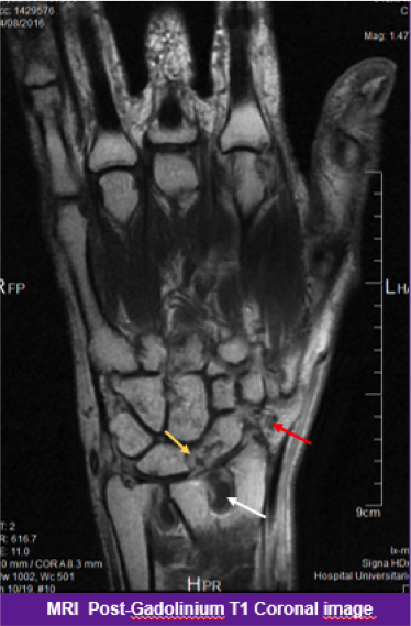
Methods: Exploratory, non-blinded cohort; from Hospital “Dr. José E. González” since Nov. 2016-Feb. 2017, (approved by Ethics and Research Committees); 60 patients assessed by rheumatologists (ACR-EULAR 2010 criteria), were divided into 3 groups: Clinically Suspicious Arthralgia (CSA), 23 (38%); Early RA (ERA), 22 (37%) and RA 15 (25%); accepted to perform MRI 1.5T, dominant hand, simple T1, T1-gadolinium and STIR (coronal and axial sections) sequences; images were assessed by experimented radiologist
Results: Female was predominant with 83% (50); mean age 42 (19-70 years); we evaluated by OMERACT-RAMIRS 1,731 wrist sites of bones and joints, obtaining lesions (synovitis, erosions, osteitis) in 56% (964); synovitis was the most prevalent 46% (445) and Triquetrum bone the most affected in all 3 groups: CSA, 87% (20/23), ERA, 91% (20/22) and RA, 93% (14/15), see Table and Figure
Conclusion: The predominant Triquetral synovitis, could suggest us, be the first morphological site to be considered in the RA evaluation when it´is clinically suspected; however further longitudinal studies are needed to conclude it
|
CSA (n=23) |
Synovitis n (%) |
Erosions n (%) |
Osteitis n (%) |
|
Triquetrum |
20 (87) |
21 (91) |
2 (9) |
|
Lunate |
11 (48) |
22 (96) |
2 (9) |
|
Capitate |
10 (44) |
21 (91) |
4 (17) |
|
TOTAL=288 (16%) |
137 (48) |
130 (45) |
21 (8) |
|
Active erosions |
30 (26) |
||
|
ERA (n=22) |
|||
|
Triquetrum |
20 (91) |
21 (96) |
12 (55) |
|
Lunate |
16 (73) |
21 (96) |
12 (55) |
|
Scaphoid |
18 (82) |
20 (91) |
10 (46) |
|
TOTAL=408 (24%) |
181 (44) |
154 (38) |
73 (18) |
|
Active erosions |
52 (44) |
||
|
RA (n=15) |
|||
|
Triquetrum |
14 (93) |
14 (93) |
11 (73) |
|
Scaphoid |
14 (93) |
14 (93) |
7 (47) |
|
Hamate |
13 (87) |
9 (60) |
6 (40) |
|
TOTAL=268 (15%) |
127 (47) |
78 (29) |
63 (24) |
|
Active erosions |
35 (30) |
To cite this abstract in AMA style:
Larios-Forte MDC, Skinner-Taylor C, Galarza-Delgado DA, Esquivel-Valerio J, Riega-Torres J, Vega-Morales D, Perez-Onofre I. Carpal Bones Affectation Frequency in Rheumatoid Arthritis By Magnetic Resonance Imaging [abstract]. Arthritis Rheumatol. 2018; 70 (suppl 9). https://acrabstracts.org/abstract/carpal-bones-affectation-frequency-in-rheumatoid-arthritis-by-magnetic-resonance-imaging/. Accessed .« Back to 2018 ACR/ARHP Annual Meeting
ACR Meeting Abstracts - https://acrabstracts.org/abstract/carpal-bones-affectation-frequency-in-rheumatoid-arthritis-by-magnetic-resonance-imaging/

