ACR Meeting Abstracts

ACR Meeting Abstracts

  • Meetings
    • ACR Convergence 2024
    • ACR Convergence 2023
    • 2023 ACR/ARP PRSYM
    • ACR Convergence 2022
    • ACR Convergence 2021
    • ACR Convergence 2020
    • 2020 ACR/ARP PRSYM
    • 2019 ACR/ARP Annual Meeting
    • 2018-2009 Meetings
    • Download Abstracts
  • Keyword Index
  • Advanced Search
  • Your Favorites
    • Favorites
    • Login
    • View and print all favorites
    • Clear all your favorites
  • ACR Meetings

Abstract Number: 0852

Bone Marrow Lesion Segmentation Using Synthetic Data and Deep Learning Models

Barak Michaely1, Ming Zhang2 and Juan Shan1, 1Pace University, New York, NY, 2Wentworth Institute of Technology, Boston, MA

Meeting: ACR Convergence 2021

Keywords: Imaging, Magnetic resonance imaging (MRI), Osteoarthritis

  • Tweet
  • Click to email a link to a friend (Opens in new window) Email
  • Click to print (Opens in new window) Print
Session Information

Date: Sunday, November 7, 2021

Title: Research Methodology Poster (0846–0854)

Session Type: Poster Session B

Session Time: 8:30AM-10:30AM

Background/Purpose: Bone marrow lesions (BML) are indicators for knee osteoarthritis (OA) and can be detected from magnetic resonance imaging (MRI). Due to the small size, low contrast, and various positions that BML may occur, it is challenging to automatically segment BMLs. Besides, not all patients with OA have BML, and for those patients with BML, not every slice has BML. The limited data becomes an obstacle to train machine learning models. In this work, we developed a novel strategy to synthesize MRI images with BML and trained deep learning models using the augmented dataset.

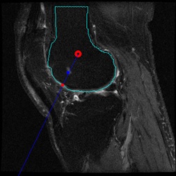
Methods: 300 cases from the Osteoarthritis Initiative (OAI) database were utilized in this study, where each case has sagittal intermediate weighted fat-suppressed (IWFS) sequence that is composed of a sequence of 36 MRI images. The BMLs in the dataset were manually delineated by domain experts as ground truth. We randomly separated the dataset into training, validation, and testing sets with 210/45/45 cases, respectively. To generate synthetic images with BML, we used images from the training set only. Three strategies were proposed. In strategy 1, for each image with BML in the training set, we selected a clean image without BML from the training set. We cropped out the BML from the original image and overlaid it on the clean image. To decide where to overlay it, we first calculated the center of the femur bone, the angle to the vertical direction, and the projected point on the bone edge in the original image, as shown in Figure 1. We also calculated the distance from the center to the lesion, and from the center to the bone edge. Then we applied the same distance percentage and same angle when overlaying the BML to the clean image. In strategy 2, everything is the same as strategy 1 except a fixed distance percentage was used to make sure the lesion is close to the bone boundary. In strategy 3, we introduced variations about the angle. A random angle from a range of -20 to +20 degrees from the vertical direction of the bone was applied when overlaying the BML. Figure 2 shows examples of the three strategies. After data generation, we first trained a deep learning model, called U-net, using the original BML images from the training set, as our baseline. Then for each strategy, a separate U-net model was trained, using the combination of original BML images plus the synthetic images.

Results: We reported the Dice coefficient and Similarity for each model in Table 1. An interesting finding is that for all the strategies, using 50% of the synthetic data produced higher accuracy than using 100% of the synthetic data. Therefore, we presented the results of using 50% of the synthetic data for each strategy. As Table 1 shows, the best accuracy was achieved by strategy 2, with more than 0.1 improvement in Dice and around 0.1 improvement in Similarity.

Conclusion: We developed an effective synthetic image generation method for BML segmentation. The accuracy was improved after adding the synthetic images to the training set. Future work could focus on refining the strategies for more realistic images and find the balanced combination between original images and synthetic images.

Figure 1. Bone center and projection on the edge (red point). BML is marked by the blue point.

Figure 2. Example images for each strategy (the highlighted arrows mark the BML location).

Table 1: BML segmentation performance on the testing set using different augmentation strategies.


Disclosures: B. Michaely, None; M. Zhang, None; J. Shan, None.

To cite this abstract in AMA style:

Michaely B, Zhang M, Shan J. Bone Marrow Lesion Segmentation Using Synthetic Data and Deep Learning Models [abstract]. Arthritis Rheumatol. 2021; 73 (suppl 9). https://acrabstracts.org/abstract/bone-marrow-lesion-segmentation-using-synthetic-data-and-deep-learning-models/. Accessed .
  • Tweet
  • Click to email a link to a friend (Opens in new window) Email
  • Click to print (Opens in new window) Print

« Back to ACR Convergence 2021

ACR Meeting Abstracts - https://acrabstracts.org/abstract/bone-marrow-lesion-segmentation-using-synthetic-data-and-deep-learning-models/

Advanced Search

Your Favorites

You can save and print a list of your favorite abstracts during your browser session by clicking the “Favorite” button at the bottom of any abstract. View your favorites »

All abstracts accepted to ACR Convergence are under media embargo once the ACR has notified presenters of their abstract’s acceptance. They may be presented at other meetings or published as manuscripts after this time but should not be discussed in non-scholarly venues or outlets. The following embargo policies are strictly enforced by the ACR.

Accepted abstracts are made available to the public online in advance of the meeting and are published in a special online supplement of our scientific journal, Arthritis & Rheumatology. Information contained in those abstracts may not be released until the abstracts appear online. In an exception to the media embargo, academic institutions, private organizations, and companies with products whose value may be influenced by information contained in an abstract may issue a press release to coincide with the availability of an ACR abstract on the ACR website. However, the ACR continues to require that information that goes beyond that contained in the abstract (e.g., discussion of the abstract done as part of editorial news coverage) is under media embargo until 10:00 AM ET on November 14, 2024. Journalists with access to embargoed information cannot release articles or editorial news coverage before this time. Editorial news coverage is considered original articles/videos developed by employed journalists to report facts, commentary, and subject matter expert quotes in a narrative form using a variety of sources (e.g., research, announcements, press releases, events, etc.).

Violation of this policy may result in the abstract being withdrawn from the meeting and other measures deemed appropriate. Authors are responsible for notifying colleagues, institutions, communications firms, and all other stakeholders related to the development or promotion of the abstract about this policy. If you have questions about the ACR abstract embargo policy, please contact ACR abstracts staff at [email protected].

Wiley

  • Online Journal
  • Privacy Policy
  • Permissions Policies
  • Cookie Preferences

© Copyright 2025 American College of Rheumatology