Session Information
Date: Sunday, November 13, 2016
Title: Imaging of Rheumatic Diseases - Poster I: Ultrasound and Emerging Technologies
Session Type: ACR Poster Session A
Session Time: 9:00AM-11:00AM
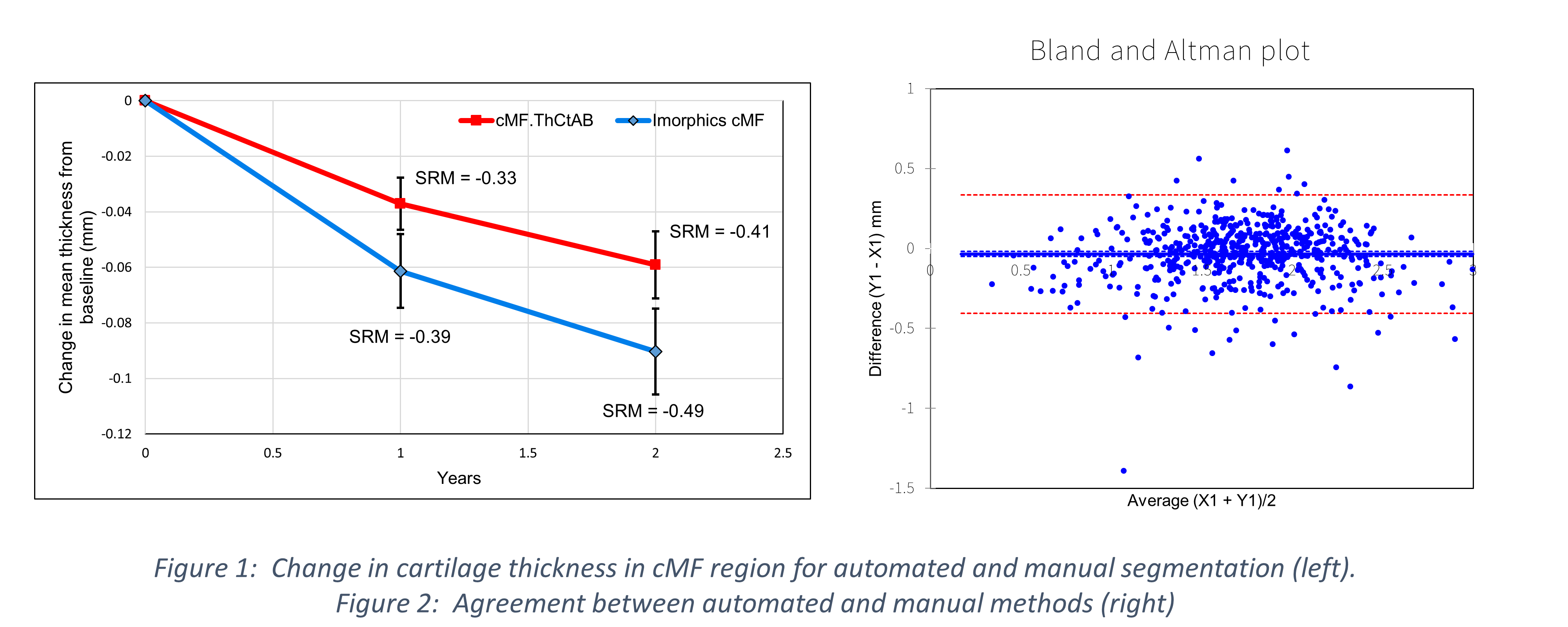
Background/Purpose: A fully automated cartilage segmentation method based on active appearance modelling (AAM), has demonstrated superior performance for a number of tissues including knee and prostate (using MRI), and abdominal, head and neck organs (using CT). Automated segmentation of tissues with minimal change is often insensitive, due to smoothing approximations of such change. In this study we compared the responsiveness of cartilage thickness in the central medial femur region (cMF) using either automatic segmentation or careful manual segmentation, using 565 knees from the Osteoarthritis Initiative over a 2-year period, together with the agreement between the 2 methods.
Methods: 565 knees with OA were analysed at 0,1, and 2 years within the OAI, and results are available on the OAI website (https://oai.epi-ucsf.org/datarelease/ImageAssessments.asp). We compared change from baseline using a pairwise student t-test 0f mean thickness of the manual cMF.ThCtAb region, and a comparable region within an AAM of the femur (Figure 1). Responsiveness was assessed using the standardised response mean (SRM). Agreement between the methods was assessed using a Bland Altman plot (Figure 2). Each image is automatically segmented using AAMs of bone and cartilage through multi-start optimisation. Initially, this fits low-density low-resolution models but ends in a robust matching of detailed high resolution models. Finally, the voxels contained in the cartilage region are assigned with a non–linear regression function, trained with a probably approximately correct (PAC) learning method.
Results: Change in manual cMF at 1 year was 0.037mm, confidence limit (0.028,0.046), p<10-4, SRM -0.33; at 2 years was 0.059 (0.047,0.081), p<10-4, SRM -0.41. Change in automated cMF at 1 years was 0.061(0.048,0.074), p<10-4, SRM -0.39; at 2 years was 0.090 (0.075,0.105), p<10-4, SRM -0.49. The methods agreed well, with a systematic bias of -0.034mm, with a 95% confidence limit of 0.37mm, comparable to manual test-retest agreement (unpublished data)
Conclusion: Automated cartilage segmentation using AAMs provides comparable cartilage thickness measures to careful manual segmentation, and improved responsiveness. Manual cartilage segmentation is labour intensive and limits the pursuit of OA clinical trials. Automation now provides an equally accurate alternative, allowing for the segmentation of large datasets such as the OAI. 
To cite this abstract in AMA style:
Guillard G, Vincent GR, Conaghan PG, Brett A, Bowes MA. Automated Segmentation of Cartilage Provides Comparable Accuracy and Better Responsiveness Than Manual Segmentation: Data from the Osteoarthritis Initiative [abstract]. Arthritis Rheumatol. 2016; 68 (suppl 10). https://acrabstracts.org/abstract/automated-segmentation-of-cartilage-provides-comparable-accuracy-and-better-responsiveness-than-manual-segmentation-data-from-the-osteoarthritis-initiative/. Accessed .« Back to 2016 ACR/ARHP Annual Meeting
ACR Meeting Abstracts - https://acrabstracts.org/abstract/automated-segmentation-of-cartilage-provides-comparable-accuracy-and-better-responsiveness-than-manual-segmentation-data-from-the-osteoarthritis-initiative/
