Session Information
Date: Sunday, November 12, 2023
Title: (0380–0422) RA – Diagnosis, Manifestations, and Outcomes Poster I
Session Type: Poster Session A
Session Time: 9:00AM-11:00AM
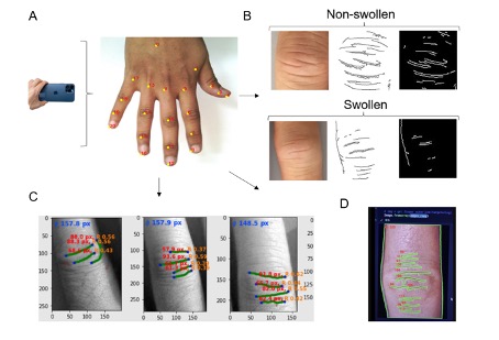
Background/Purpose: We have previously shown that automated detection and processing of dorsal finger fold patterns from hand photographs can be used as a digital biomarker for joint swelling in rheumatoid arthritis.
In this study, we tested different computer vision and deep learning methods for the automated quantification of dorsal finger folds and diameter of proximal interphalangeal finger joints (PIP) in patients with rheumatoid arthritis. Additionally, the selected model aimed to evaluate the difference in PIP finger joint swelling between healthy individuals and rheumatoid arthritis patients.
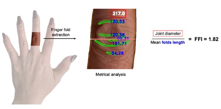
Methods: We evaluated the detection and measurement of PIP joint diameter and dorsal finger fold length on hand photographs in 1783 joints of patients with rheumatoid arthritis by canny edge or ridge detection computer vision models or a newly trained convolutional neural network, respectively. The models have been trained to calculate the finger fold index (FFI, potential biomarker for joint swelling), defined as the ratio between pixel length of joint diameter and mean recognized finger folds. In an independent healthy control dataset, the FFI has been calculated to be compared with rheumatoid arthritis PIP joints.
Results: Canny edge and ridge detection were suitable models to detect dorsal finger fold patterns. The prediction of joint diameter and finger fold pixel length was best achieved in a newly trained deep neural network model, where the FFI was predictable in 93% of the images. The accuracy of correct detection of joint diameter measurement was 91%. In 1783 PIP joints of patients with rheumatoid arthritis, the mean FFI was significantly higher than in 168 healthy controls PIP joints (3.42 ± SD 1.04 and 2.15 ± SD 0.68, respectively, p-value < 0.05).
Conclusion: Finger fold index calculated by a deep neural network model seems a reliable tool for the metrical analysis and thus gradeless detection of swelling in patients with rheumatoid arthritis. Standard deviation for FFI in patients with rheumatoid arthritis was higher than in healthy patients. Therefore, further investigation by stratifying patients into different disease activity levels as well as the correlation of FFI with longitudinal changes of clinical scores on a joint-level and general disease activity is ongoing.
To cite this abstract in AMA style:
Blanchard M, Maglione J, Koller C, Hermann P, Brüschweiler D, Kleyer A, Hügle T. Automated Detection and Quantification of Hand Joint Swelling in Rheumatoid Arthritis: Computer Vision, Deep Neural Network Models, and a Potential Biomarker for Disease Activity Monitoring [abstract]. Arthritis Rheumatol. 2023; 75 (suppl 9). https://acrabstracts.org/abstract/automated-detection-and-quantification-of-hand-joint-swelling-in-rheumatoid-arthritis-computer-vision-deep-neural-network-models-and-a-potential-biomarker-for-disease-activity-monitoring/. Accessed .« Back to ACR Convergence 2023
ACR Meeting Abstracts - https://acrabstracts.org/abstract/automated-detection-and-quantification-of-hand-joint-swelling-in-rheumatoid-arthritis-computer-vision-deep-neural-network-models-and-a-potential-biomarker-for-disease-activity-monitoring/


