Session Information
Date: Sunday, October 21, 2018
Title: Pediatric Rheumatology – Clinical Poster I: Lupus, Sjögren’s Disease, and Myositis
Session Type: ACR Poster Session A
Session Time: 9:00AM-11:00AM
Background/Purpose:
There is an unmet need for more objective disease outcome measures in Juvenile Myositis (JM) patients. This pilot study sought to test the reliability, validity and responsiveness of muscle ultrasound (US) modalities as outcome measures in JM subjects.
Methods:
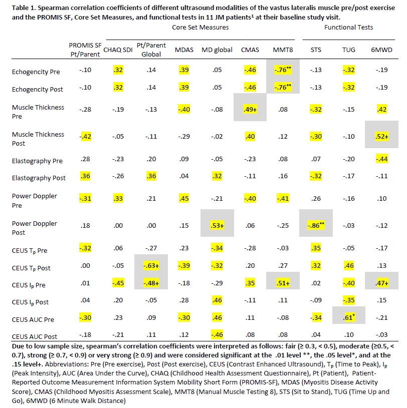
This prospective, consecutive observational JM cohort (using Bohan and Peter criteria) had clinical, functional and muscle US data collected at baseline, 3 and 6 months. US modalities assessing muscle consistency and perfusion [i.e. Gray Scale with Echogenicity and Muscle Thickness, Power Doppler, 2D Shear wave© Elastography, and Contrast Enhanced US with Lumason© (CEUS)] were performed unilaterally on the proximal vastus lateralis (VL) at each study visit before and after all exercises including functional measure tests. Spearman correlation was utilized to evaluate the relationship of baseline US VL data pre- and post-exercise to International Myositis Assessment and Clinical Studies Group (IMACS) validated Core Set Measures (CSM). Descriptive statistics for VL US longitudinal measurements were performed and graphed to visualize trends. Subjects were characterized as change vs no change, guided by ACR/EULAR myositis response criteria and designated as ‘active’ or ‘inactive’ disease (PRINTO definition and physician judgement).
Results:
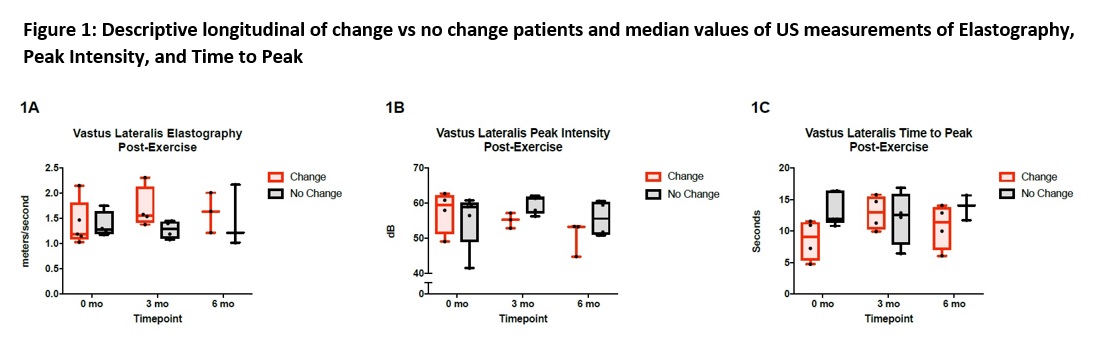
Eleven enrolled subjects included primarily Caucasian females (mean age 10.7 ± 4.2 years) with a mean disease duration of 36 ± 23.7 months. At baseline, several US measures had fair to strong correlations (rs > 0.30) with CSM, with most associations being stronger post-exercise (Table 1). Longitudinal assessment confirmed this post-exercise trend with disease status. The ‘no change’ subjects demonstrate consistent muscle US values over time (reliability) (Fig1). Patients with initial ‘active’ disease showed an increase in elastography following exercise, reflecting an increase in muscle stiffness (Fig1A), and a decrease in degree of perfusion post exercise, measured by Peak Intensity CEUS, over time (Fig 1B). Additionally, the speed of perfusion on Time to Peak CEUS increased in subjects who clinically improved in their disease status (change group) (Fig1C).
Conclusion:
Following exercise in active JM patients, the VL remains stiffer with less capillary enhancement compared to stable patients, likely reflecting residual vasculopathy. In patients showing clinical improvement, speed of perfusion (Tp CEUS) increases over time and may be useful as a future treatment response measure. Further longitudinal analysis is underway with healthy controls to strengthen these findings.
To cite this abstract in AMA style:
Tasan L, Brunner E, Squires J, Aggarwal R, Oddis CV, Zigler CK, Schollaert-Fitch K, Mirizio E, Torok KS. Analyze Myositis with Ultrasound and Exercise (AMUSE) Kids- Initial Analysis of Longitudinal Data [abstract]. Arthritis Rheumatol. 2018; 70 (suppl 9). https://acrabstracts.org/abstract/analyze-myositis-with-ultrasound-and-exercise-amuse-kids-initial-analysis-of-longitudinal-data/. Accessed .« Back to 2018 ACR/ARHP Annual Meeting
ACR Meeting Abstracts - https://acrabstracts.org/abstract/analyze-myositis-with-ultrasound-and-exercise-amuse-kids-initial-analysis-of-longitudinal-data/