Session Information
Date: Tuesday, October 28, 2025
Title: (2377–2436) Systemic Lupus Erythematosus – Diagnosis, Manifestations, & Outcomes Poster III
Session Type: Poster Session C
Session Time: 10:30AM-12:30PM
Background/Purpose: Fluorine-18 fluorodeoxyglucose positron emission tomography/computed tomography (18F-FDG PET/CT) is a noninvasive imaging modality widely used to assess inflammatory activity in various autoimmune and hematologic disorders. The bone marrow (BM), as the site of hematopoiesis, may play a key role in the pathogenesis of systemic lupus erythematosus (SLE). This study aimed to investigate whether BM metabolic activity on PET/CT is associated with hematologic manifestations and to assess its potential as a predictive biomarker in SLE.
Methods: We retrospectively analyzed patients with SLE who underwent 18F-FDG PET/CT imaging at baseline and had available follow-up data for at least 6 months. Patients with malignancy were excluded. BM FDG uptake was quantified using standardized uptake values (SUV), and the BM-to-liver ratio (BLR) was calculated using both SUVmax and SUVmean. Hematologic manifestations were defined as leukopenia, anemia, and/or thrombocytopenia. Associations between BLR indices and hematologic involvement were assessed using Pearson correlation and multivariable logistic regression adjusted for age, sex, and comorbidities. Receiver operating characteristic (ROC) curve analysis was conducted to evaluate predictive performance.
Results: Among 76 patients with SLE (median age 48.0 years; 86.0% female), 21 (27.6%) exhibited baseline hematologic abnormalities. Median BM SUVmax and SUVmean were 2.40 (SD, 0.65) and 1.65 (SD, 0.50), respectively. Median BLRmax and BLRmean were 1.58 (SD, 0.52) and 1.10 (SD, 0.41), respectively. Both BLRmax and BLRmean were positively correlated with baseline hematologic involvement (r = 0.282, p = 0.014 and r = 0.264, p = 0.021, respectively). Elevated baseline BLR values were independently associated with the development of hematologic abnormalities at 6 months (adjusted ORs: 1.57 [95% CI: 1.15–2.13] for BLRmax and 1.42 [1.10–1.83] for BLRmean; both p < 0.01). ROC curve analysis yielded an AUC of 0.76 for BLRmax in predicting future cytopenia, with an optimal cut-off value of 1.8 (sensitivity 78%, specificity 72%).
Conclusion: Increased BM-metabolic activity relative to the liver on PET/CT is associated with both initial and subsequent hematologic manifestations in patients with SLE. BLR, particularly BLRmax, may serve as a noninvasive imaging biomarker for predicting hematologic involvement, underscoring a potential role of the BM in SLE pathophysiology.
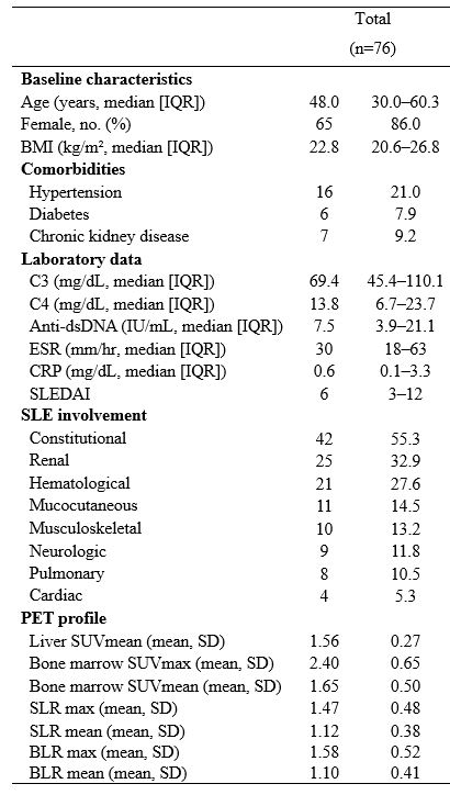 Baseline characteristics of patients with systemic lupus erythematosus who underwent 18F-FDG PET/CT.
To cite this abstract in AMA style:
Ko S, Kim Y, Yoon S, Ahn S, Oh J, Kim Y, Lee C, Yoo B, Lee D, Hong S. Bone marrow- activity on 18F-FDG PET/CT as a Predictor of Hematologic Manifestations in Systemic Lupus Erythematosus [abstract]. Arthritis Rheumatol. 2025; 77 (suppl 9). https://acrabstracts.org/abstract/bone-marrow-activity-on-18f-fdg-pet-ct-as-a-predictor-of-hematologic-manifestations-in-systemic-lupus-erythematosus/. Accessed .« Back to ACR Convergence 2025
ACR Meeting Abstracts - https://acrabstracts.org/abstract/bone-marrow-activity-on-18f-fdg-pet-ct-as-a-predictor-of-hematologic-manifestations-in-systemic-lupus-erythematosus/