ACR Meeting Abstracts

ACR Meeting Abstracts

  • Meetings
    • ACR Convergence 2025
    • ACR Convergence 2024
    • ACR Convergence 2023
    • 2023 ACR/ARP PRSYM
    • ACR Convergence 2022
    • ACR Convergence 2021
    • 2020-2009 Meetings
    • Download Abstracts
  • Keyword Index
  • Advanced Search
  • Your Favorites
    • Favorites
    • Login
    • View and print all favorites
    • Clear all your favorites
  • ACR Meetings

Abstract Number: 1964

Performance of Artificial Intelligence-Based Salivary Gland Ultrasound in Sjögren Syndrome

Otto Olivas1, Mónica García-Sevilla2, Lucía Cubero2, Javier Pascau2 and Esperanza Naredo3, 1Department of Rheumatology and Joint and Bone Research Unit. Fundación Jiménez Díaz University Hospital and Health Research Institute FJD-UAM, Madrid, Spain, Madrid, Madrid, Spain, 2Department of Bioengineering, Carlos III University of Madrid, Leganes, Madrid, Spain and Health Research Institute Gregorio Marañón, Madrid, Spain, Madrid, Madrid, Spain, 3Department of Rheumatology and Joint and Bone Research Unit. Fundación Jiménez Díaz University Hospital and Health Research Institute FJD-UAM, Madrid, Spain, MADRID, Spain

Meeting: ACR Convergence 2025

Keywords: Imaging, Sjögren's syndrome, Ultrasound

  • Tweet
  • Click to email a link to a friend (Opens in new window) Email
  • Click to print (Opens in new window) Print
Session Information

Date: Tuesday, October 28, 2025

Title: (1936–1971) Imaging of Rheumatic Diseases Poster

Session Type: Poster Session C

Session Time: 10:30AM-12:30PM

Background/Purpose: To develop a deep learning model for classifying ultrasound images of salivary glands (SG) [parotid (PG) and submandibular (SMG)] based on the Outcome Measures in Rheumatology (OMERACT) 0-3 semiquantitative scoring system for B-mode ultrasound-assessed parenchymal abnormalities in SG in Sjögren syndrome (SjS) (Figure 1)

Methods: For training, we used 3 datasets of SG (PG and SMG) ultrasound images representative of the OMERACT scores: 1) 225 images from 150 suspected or confirmed SjS patients acquired with different ultrasound systems across 4 European centers (https://www.frontiersin.org/articles/10.3389/fmed.2020.581248/full); 2) 80 images from 20 SjS patients provided by a European center; 3) Of 39 SjS patients and 10 controls, 6 images of each acquired by 2 experts with 2 systems (a high-end and a wireless handheld device) in a European center (1176 images). We used a ResNet18 architecture, which was pre-trained on ImageNet and then fine-tuned for this task. The model was trained hierarchically: initially classifying broad categories (scores 0/1 vs. 2/3) and subsequently refining the classification for the specific stages of the disease. 70% of the images were used for training, 15% for validation, and 15% for test. For training, we used a dataset of ultrasound images along with labels indicating the grade of SG involvement. We employed standard deep learning techniques such as data augmentation (e.g., random flipping and brightness adjustments) to increase the model’s robustness and generalization capabilities and a step learning rate scheduler to optimize the training process. Precision (correct classifications per predicted class), recall (correct classifications per actual class), and F1-score (harmonic mean of precision and recall) were calculated for each grading class.

Results: The overall performance of the model for grading classes is shown in Table 1. The broad model achieved an accuracy of 90% in distinguishing between 0/1 and 2/3 grades. However, the more specific models, fine-tuned for the 0/1 and 2/3 categories, had lower accuracy (approximately 65% for each). This reflects the increased difficulty of fine-grained classification. The confusion matrix (Figure 2) showed that the model performed well in classifying SG with grade 3 but faced challenges with intermediate categories (class 1 and class 2). The network showed difficulty discriminating between grade 0 and 1; on retrospective review of the ultrasound labels, we noted that this boundary is visually subtle and prone to greater inter-observer variability, which likely contributes to the model’s confusion between these two lowest scores.

Conclusion: Our results showed the potential of deep learning models, specifically convolutional neural networks, to classify SG ultrasound images based on parenchymal involvement, offering a promising tool for SjS diagnosis. While the broad classification model performed very well, the fine-tuned models, which differentiate between more specific disease stages, still faced challenges. Future work should refine the fine-tuned models with additional data, allowing more complex architecture or domain-specific adjustments.
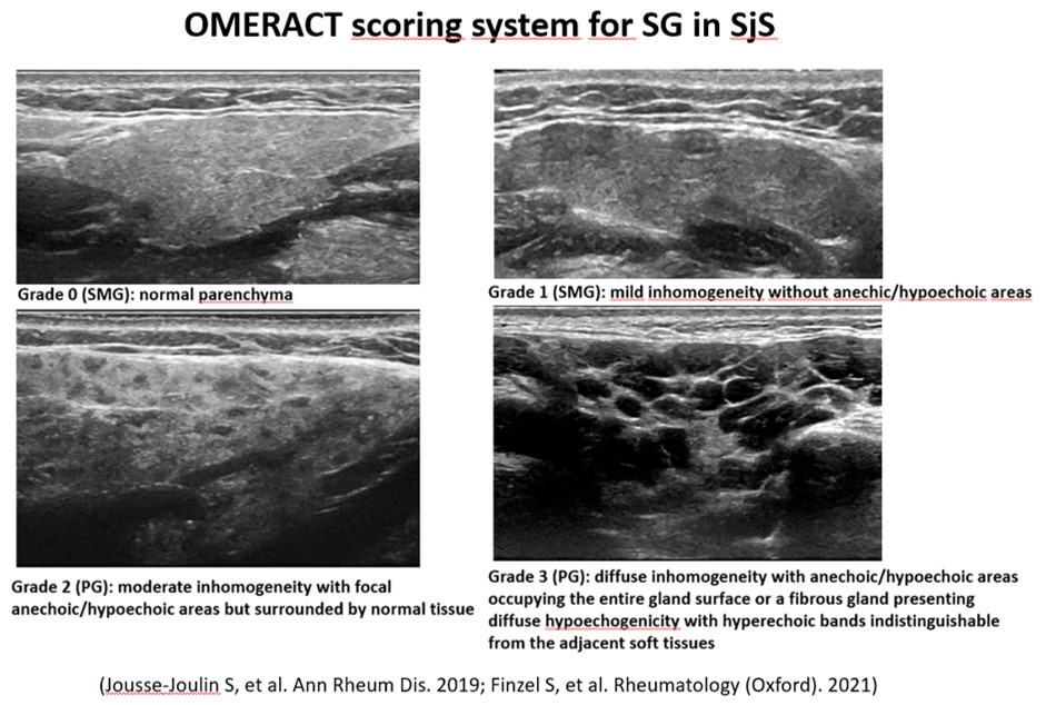
Supporting image 1Figure 1

Supporting image 2Figure 2

Supporting image 3Table 1


Disclosures: O. Olivas: None; M. García-Sevilla: None; L. Cubero: None; J. Pascau: None; E. Naredo: None.

To cite this abstract in AMA style:

Olivas O, García-Sevilla M, Cubero L, Pascau J, Naredo E. Performance of Artificial Intelligence-Based Salivary Gland Ultrasound in Sjögren Syndrome [abstract]. Arthritis Rheumatol. 2025; 77 (suppl 9). https://acrabstracts.org/abstract/performance-of-artificial-intelligence-based-salivary-gland-ultrasound-in-sjogren-syndrome/. Accessed .
  • Tweet
  • Click to email a link to a friend (Opens in new window) Email
  • Click to print (Opens in new window) Print

« Back to ACR Convergence 2025

ACR Meeting Abstracts - https://acrabstracts.org/abstract/performance-of-artificial-intelligence-based-salivary-gland-ultrasound-in-sjogren-syndrome/

Advanced Search

Your Favorites

You can save and print a list of your favorite abstracts during your browser session by clicking the “Favorite” button at the bottom of any abstract. View your favorites »

Embargo Policy

All abstracts accepted to ACR Convergence are under media embargo once the ACR has notified presenters of their abstract’s acceptance. They may be presented at other meetings or published as manuscripts after this time but should not be discussed in non-scholarly venues or outlets. The following embargo policies are strictly enforced by the ACR.

Accepted abstracts are made available to the public online in advance of the meeting and are published in a special online supplement of our scientific journal, Arthritis & Rheumatology. Information contained in those abstracts may not be released until the abstracts appear online. In an exception to the media embargo, academic institutions, private organizations, and companies with products whose value may be influenced by information contained in an abstract may issue a press release to coincide with the availability of an ACR abstract on the ACR website. However, the ACR continues to require that information that goes beyond that contained in the abstract (e.g., discussion of the abstract done as part of editorial news coverage) is under media embargo until 10:00 AM CT on October 25. Journalists with access to embargoed information cannot release articles or editorial news coverage before this time. Editorial news coverage is considered original articles/videos developed by employed journalists to report facts, commentary, and subject matter expert quotes in a narrative form using a variety of sources (e.g., research, announcements, press releases, events, etc.).

Violation of this policy may result in the abstract being withdrawn from the meeting and other measures deemed appropriate. Authors are responsible for notifying colleagues, institutions, communications firms, and all other stakeholders related to the development or promotion of the abstract about this policy. If you have questions about the ACR abstract embargo policy, please contact ACR abstracts staff at [email protected].

Wiley

  • Online Journal
  • Privacy Policy
  • Permissions Policies
  • Cookie Preferences

© Copyright 2025 American College of Rheumatology