Session Information
Session Type: Poster Session C
Session Time: 10:30AM-12:30PM
Background/Purpose: Interstitial lung disease (ILD) is the leading cause of death in MDA5 dermatomyositis (MDA5-DM). Fibrosis and inflammation are pivotal drivers in the pathogenesis of DM-ILD. This study aims to investigate the imaging characteristics of ⁶⁸Ga-FAPI-04 and ¹⁸F-FDG PET/CT in MDA5 DM ILD, and to evaluate the association between pulmonary metabolic parameters of fibrosis and inflammation and mortality in MDA5 DM-ILD.
Methods: Between August 2022 and November 2024, 20 patients with newly diagnosed (duration ≤6 months) MDA5 DM were enrolled in the study, followed by a 6-month longitudinal observation period.All participants underwent baseline ⁶⁸Ga-FAPI-04 PET/CT and ¹⁸F-FDG PET/CT scanning. Parameters including total lesion volume (TLV), total target-to-background ratio (TBR_total), mean TBR (TBR_mean), and maximum TBR (TBR_max) based on standardized uptake values (SUV) were quantified.Non-parametric Mann-Whitney U tests compared group differences.Spearman’s rank correlation analyes parameter associations, and point-biserial correlation coefficients quantified associations with mortality.
Results: The study included 20 patients with a mean age of 50.2 ± 13.8 years ,and 60% were male. The median disease duration was 2.0 months (interquartile range [IQR], 1.0–3.0 months). During follow-up , mortality was observed in 3 of the 20 enrolled patients. Lesion volumes quantified by ⁶⁸Ga-FAPI-04 and ¹⁸F-FDG PET/CT exhibited high concordance, with a robust positive correlation (Spearman’s ρ = 0.93), indicating spatial colocalization of inflammation and fibroblast activation in the affected regions. TBRmax FAPI and TBRmean also showed a strong positive correlation (Spearman’s ρ=0.92). Deceased patients demonstrated significantly elevated ¹⁸F-FDG TBRmax values compared to survivors (median: 5.97 vs. 2.04, P=0.04), suggesting that inflammatory activity within lung lesions may contribute to the mortality risk of individuals with MDA5 DM.
Conclusion: In MDA5 DM patients, pulmonary lesions were characterized by hypermetabolism and active fibrosis. The ¹⁸F-FDG TBRmax emerged as a potential prognostic marker for mortality, reflecting localized hypermetabolism in lung tissue that may indicate aggressive disease progression and correlate with poor clinical outcomes. These findings highlight the complementary roles of inflammation (FDG) and fibrosis (FAPI) imaging in characterizing disease severity. Further studies are warranted to validate the prognostic utility of TBRmax FDG and explore integrated imaging strategies for risk stratification in MDA5 DM-associated interstitial lung disease.
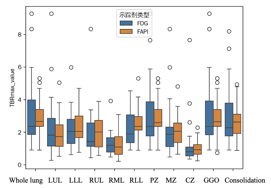 Figure 1 68Ga-FAPI-04 and 18F-FDG PET/CT imaging in a representative patient
.jpg) Figure 2 The association between pulmonary metabolic parameters of FAPI and FDG in MDA5 DM-ILD.
Figure 2 The association between pulmonary metabolic parameters of FAPI and FDG in MDA5 DM-ILD.
.jpg) Figure 3 Boxplot of TBRmax for FDG and FAPI Tracers in Different Regions
Figure 3 Boxplot of TBRmax for FDG and FAPI Tracers in Different Regions
To cite this abstract in AMA style:
Li J, Wei W, Li L, Xu W, Liu J, Ye S. 68Ga-FAPI-04 and 18F-FDG PET/ CT in MDA5 dermatomyositis-associated interstitial lung disease: a single-center, pilot study [abstract]. Arthritis Rheumatol. 2025; 77 (suppl 9). https://acrabstracts.org/abstract/68ga-fapi-04-and-18f-fdg-pet-ct-in-mda5-dermatomyositis-associated-interstitial-lung-disease-a-single-center-pilot-study/. Accessed .« Back to ACR Convergence 2025
ACR Meeting Abstracts - https://acrabstracts.org/abstract/68ga-fapi-04-and-18f-fdg-pet-ct-in-mda5-dermatomyositis-associated-interstitial-lung-disease-a-single-center-pilot-study/