Session Information
Session Type: Poster Session C
Session Time: 10:30AM-12:30PM
Background/Purpose: Nailfold capillaroscopy plays a critical role in the diagnosis and management of systemic sclerosis (SSc) and related connective tissue diseases. However, access to capillaroscopy remains limited outside specialized rheumatology centers. Dermatoscopy is a more accessible alternative, but its use for nailfold evaluation lacks standardization. In 2024, we demonstrated preliminary evidence that artificial intelligence (AI) could support dermatoscopic nailfold analysis in rheumatology clinics (Maldonado et al., Arthritis Rheumatol. 2024). Building on that work, we now present a refined and more efficient AI model designed for possible real-world implementation.
Methods: We developed a multi-label image classification model using 1,405 dermatoscopic nailfold images labeled for five key features: abnormal shapes, giant capillaries (replacing the broader “enlarged” category for better dermatoscopic specificity), hemorrhages, low capillary density, and image blurriness. Unlike our 2024 approach, which used separate binary models per feature, this year’s model employs a unified architecture to predict all labels simultaneously—enhancing computational efficiency and deployment feasibility. The model was based on ConvNeXt V2 and trained on an 80/20 stratified split, with performance evaluated using standard machine learning metrics. Images were retrieved using a 200x magnification lens adapted for smartphones (value $30).
Results: The model achieved strong performance across all target features (table 1), with F1-scores as follows: abnormal shapes (0.643), giant capillaries (0.647), hemorrhages (0.649), low density (0.667), and image too blurry (0.874), yielding a mean F1-score of 0.696. The multi-label framework significantly reduced inference time and memory usage while maintaining consistent accuracy across folds (table 2). The model also demonstrated robustness despite the lower resolution typical of dermatoscopic images compared to nailfold capillaroscopy.
Conclusion: This AI-driven model shows promising accuracy in detecting microvascular abnormalities from dermatoscopic images and represents a meaningful step toward accessible, point-of-care screening for SSc and related diseases. By leveraging the lightweight ConvNeXt V2 architecture in a multi-label framework, we pave the way for integration into mobile applications using a smartphone camera and dermatoscopy lens. Such a tool could support early detection of microvascular abnormalities in patients with Raynaud’s phenomenon, especially in non-specialist or resource-limited settings. However, given the high proportion of low-quality (blurry) images in dermatoscopy and in alignment with current recommendations (Mislav R. et al. Clin Exp Rheumatol. 2020;38 Suppl 125:132–136), nailfold video capillaroscopy should be pursued for comprehensive evaluation when clinically indicated. Prospective validation and clinical integration studies are ongoing.
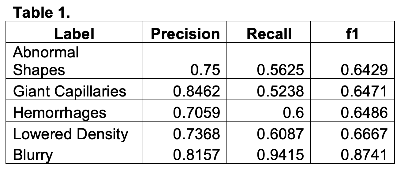 Table 1. Model performance
.jpg) Table 2. Sample of images by label.
Table 2. Sample of images by label.
To cite this abstract in AMA style:
Maldonado G, Ramos ibáñez E, Gracia Tello B, Frech T. Revolutionizing Microvascular Screening: AI-Powered Dermatoscopy for Efficient Nailfold Capillary Evaluation [abstract]. Arthritis Rheumatol. 2025; 77 (suppl 9). https://acrabstracts.org/abstract/revolutionizing-microvascular-screening-ai-powered-dermatoscopy-for-efficient-nailfold-capillary-evaluation/. Accessed .« Back to ACR Convergence 2025
ACR Meeting Abstracts - https://acrabstracts.org/abstract/revolutionizing-microvascular-screening-ai-powered-dermatoscopy-for-efficient-nailfold-capillary-evaluation/