Session Information
Date: Tuesday, October 28, 2025
Title: (1830–1854) Systemic Lupus Erythematosus – Etiology and Pathogenesis Poster
Session Type: Poster Session C
Session Time: 10:30AM-12:30PM
Background/Purpose: Lupus nephritis (LN) occurs in over 50% of patients with pediatric systemic lupus erythematosus (pSLE) and results in significant morbidity due to suboptimal kidney remission rates and the sequelae of prolonged intensive immunosuppressive therapy. LN is often patchy, with some glomeruli severely damaged while others remain histologically unaffected in the same kidney. However, the local factors that drive this patchy tissue injury remain unclear. Using novel spatial transcriptomic technology, we interrogated microanatomic transcriptional differences between histologically damaged and unaffected glomeruli and adjacent tubules in pSLE LN to understand local drivers of renal injury.
Methods: Archived pre-treatment FFPE pediatric renal biopsies were stained with H&E. Using the Visium 10x platform, spatially barcoded gene expression libraries mapped to 55μM diameter spots were sequenced. A pediatric pathologist identified histologically damaged and unaffected glomeruli based on H&E staining. Loupe Browser (10X Genomics) was used to annotate damaged versus unaffected glomeruli (Figure 1A) and adjacent tubules (Figure 2A) and their associated barcoded spots. Differential gene expression analysis of spots was performed in R. 3 cases of pSLE Class III LN, 3 cases of pSLE Class V LN, and 3 “healthy control” kidneys resected for non-immune pathology were assessed. Immunofluorescence staining was used to assess cellular localization of specific transcriptional changes at the protein level.
Results: Differential gene expression analysis revealed a unique transcriptional signature in histologically damaged compared to unaffected Class III LN glomeruli (Figure 1B). While interferon-stimulated genes were unchanged (Figure 1C), damaged Class III LN glomeruli showed increased expression of C5AR1 and both ITGA11 and FN1 compared to unaffected Class III LN glomeruli (Figure 1D) implicating complement component 5a activity and fibrosis respectively. C5AR1 expression is likely driven by myeloid cells based on analysis of single cell RNA sequencing data from the Accelerating Medicines Partnership (Figure 1E) and immunofluorescence staining (Figure 1F). Periglomerular tubules next to damaged Class III LN glomeruli show increased early complement gene expression and expression of the fibrosis inducing gene POSTN compared to those next to unaffected Class III LN glomeruli and those far from glomeruli (Figure 2B,C). All changes remain significant even after correcting for patient level differences. POSTN expression correlates with other fibrosis-related genes including LGALS1 (Figure 2D).
Conclusion: LN is characterized by a specific microanatomic patterning of inflammation and fibrosis. Damaged glomeruli and adjacent tubules in Class III LN are marked by local complement gene and C5AR1 expression and fibrosis-related genes. These results suggest novel useful biomarkers and potential therapeutic targets for LN (eg. avacopan to target C5aR and early targeting of fibrosis-related pathways). These data will inform mechanistic studies using animal models, and translational clinical studies of these novel targets.
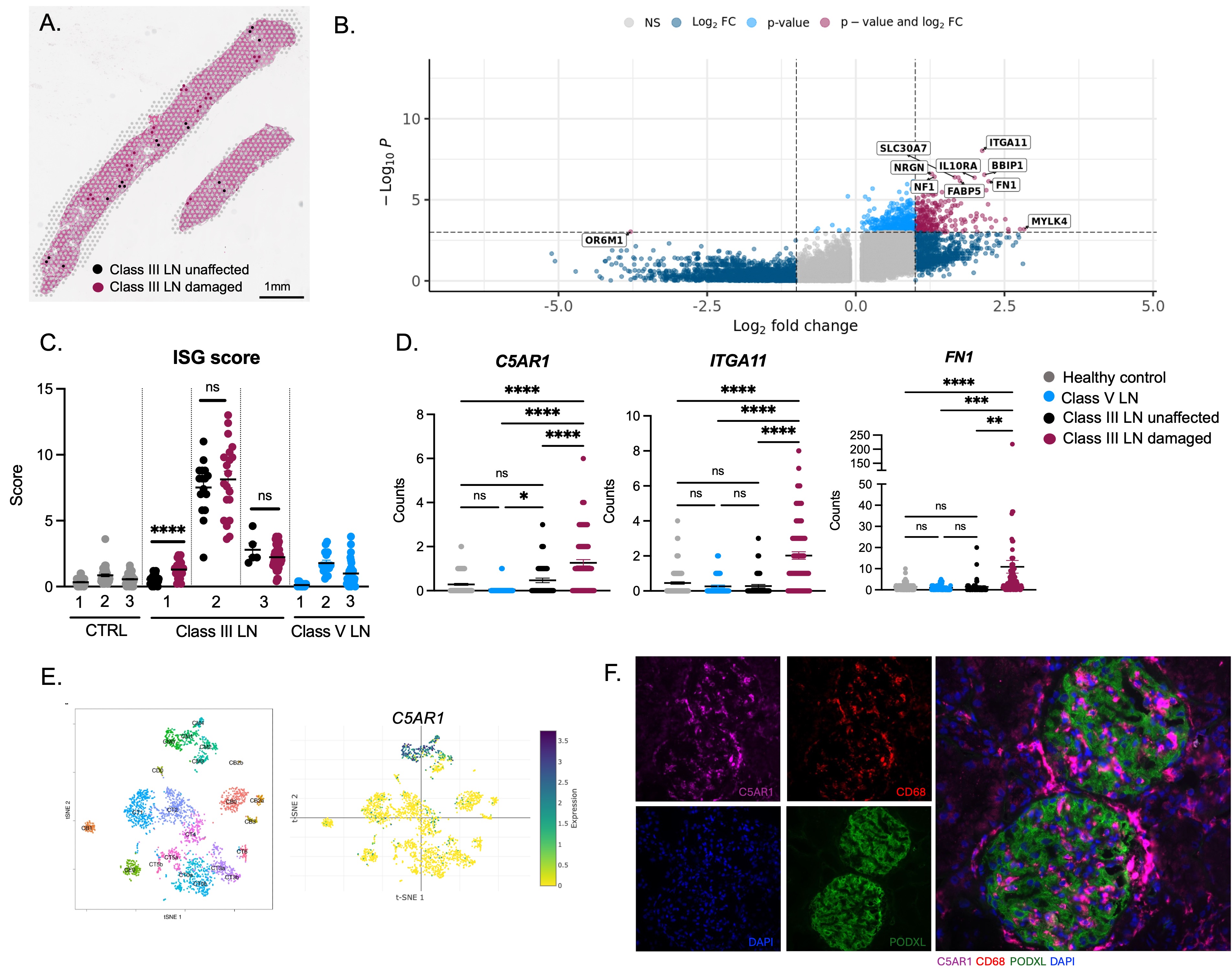 Figure 1 – Damaged Class III LN glomeruli show upregulation of C5AR1 and genes linked to fibrosis.
Spatial transcriptomics using the Visium 10x platform was performed on FFPE renal biopsies from pediatric patients with Class III LN (Nf3), Class V LN (Nf3), and “healthy” controls (Nf3). (A) Representative H&E section from Class III LN biopsy with overlay of 55uM spots showing pathologist annotation of histologically damaged (pink) and unaffected (black) glomeruli. (B) Enhanced volcano plot showing differentially expressed genes comparing histologically damaged to unaffected glomeruli Class III LN glomeruli. Positive log2 fold changes are genes with increased expression in damaged compared to unaffected glomeruli. (C) ISG score computed as the average of the expression of IFI44, IFI44L, MX1, IFIT1, HERC6. Each dot is expression from a 55uM spot. X axis numbers represent individual patients in each disease subset. **** p < 0.0001 unpaired T-test for comparison between damaged and unaffected Class III LN glomeruli. (D) C5AR1, ITGA11, FN1 expression. Each dot is expression from a 55uM spot. Nf3 healthy control. Nf3 Class III LN. Nf3 Class V LN. ****p < 0.0001, ***p < 0.001, **p < 0.01, one way ANOVA with Tukey’s post-test for multiple comparisons. (E) Publicly available single cell sequencing data from LN biopsies from the Accelerating Medicines Partnership (AMP) showing C5AR1 expression is highest in the macrophage clusters. (F) Immunofluorescence staining of C5AR1, CD68 (marks myeloid cells), PODXL (marks podocytes) in a representative Class III LN biopsy.
.jpg) Figure 2 – Transcriptional differences between periglomerular tubules close to damaged and unaffected glomeruli in Class III pSLE GN. (A) Representative H&E section from Class III LN biopsy with overlay of 55uM spots showing annotation of tubules that are periglomerular (black = close to unaffected glomeruli, pink = close to damaged glomeruli) or far from glomeruli (grey). (B) Complement gene expression in periglomerular tubules close to damaged and unaffected glomeruli. C) POSTN expression in periglomerular tubules. (D) Pearson correlation analysis showing top genes correlated with POSTN. LGALS1 expression in tubules close to damaged glomeruli and far from damaged glomeruli. Each dot represents expression from a 55uM spot. Nf3 pSLE Class III LN patients. P values are unpaired T-tests. ****p < 0.0001, ***p < 0.001, **p < 0.01, *p < 0.05.
Figure 2 – Transcriptional differences between periglomerular tubules close to damaged and unaffected glomeruli in Class III pSLE GN. (A) Representative H&E section from Class III LN biopsy with overlay of 55uM spots showing annotation of tubules that are periglomerular (black = close to unaffected glomeruli, pink = close to damaged glomeruli) or far from glomeruli (grey). (B) Complement gene expression in periglomerular tubules close to damaged and unaffected glomeruli. C) POSTN expression in periglomerular tubules. (D) Pearson correlation analysis showing top genes correlated with POSTN. LGALS1 expression in tubules close to damaged glomeruli and far from damaged glomeruli. Each dot represents expression from a 55uM spot. Nf3 pSLE Class III LN patients. P values are unpaired T-tests. ****p < 0.0001, ***p < 0.001, **p < 0.01, *p < 0.05.
To cite this abstract in AMA style:
McCuaig S, Rood J, Elliott E, Kreiger P, Behrens E. Spatial transcriptomics reveals a complex microanatomic patterning of complement mediated inflammation and fibrosis in Class III pediatric lupus nephritis associated with local histologic injury [abstract]. Arthritis Rheumatol. 2025; 77 (suppl 9). https://acrabstracts.org/abstract/spatial-transcriptomics-reveals-a-complex-microanatomic-patterning-of-complement-mediated-inflammation-and-fibrosis-in-class-iii-pediatric-lupus-nephritis-associated-with-local-histologic-injury/. Accessed .« Back to ACR Convergence 2025
ACR Meeting Abstracts - https://acrabstracts.org/abstract/spatial-transcriptomics-reveals-a-complex-microanatomic-patterning-of-complement-mediated-inflammation-and-fibrosis-in-class-iii-pediatric-lupus-nephritis-associated-with-local-histologic-injury/