Session Information
Date: Monday, October 27, 2025
Session Type: Abstract Session
Session Time: 3:00PM-3:15PM
Background/Purpose: Enthesitis affects 30–40% of PsA patients but remains difficult to diagnose due to overlap with non-specific entheseal pain. While OMERACT has standardized ultrasound (US) elementary lesions, no validated definition of inflammatory enthesitis (IE) exists at the single-enthesis level. This limits diagnostic specificity and contributes to misclassification and overtreatment. We aimed to develop an expert- and data-driven definition of sonographic IE at the single-enthesis level in PsA, with the goal of improving diagnostic accuracy and patient selection for clinical research.
Methods: Ten expert sonographers from the DUET (Diagnostic Ultrasound Enthesitis Tool)1 study independently reviewed 90 US video scans of PsA patients at six entheseal sites. Each scan was score from –10 (“Definite not IE”) to +10 (“Definite IE”), and those with ≥70% agreement and scores of +7 were labeled “Definite IE.” Experts annotated key sonographic features influencing their assessments. Quantitative analysis included group comparison of elementary lesion distribution using the DUET consensus scoring for descriptive statistics and chi-square tests. Thematic analysis was performed on free-text annotations. This data was used to inform a proposed definition of IE, which was subsequently approved through a Delphi vote.
Results: Of 90 scans, 29 (32.2%) were “Definite IE”, 21 (23.3%) “Definite not IE”, and 40 (44.5%) “Uncertain”. Most “Definite IE” had ≥4 elementary lesions, while “Uncertain” scans commonly had 2 to 4 lesions (Figure 1A). The absence of elementary lesions or presence of isolated findings characterized the “Definite not IE” group.Power Doppler (PD) grade ≥ 2 near the bone cortex was present in 97% of the “Definite IE” group and in 55% of “Uncertain” scans (Figure 1B, Figure 2F). Importantly, grades 2-3 PD were almost exclusively found in these two groups, with grade 3 almost only found in the “Definite IE” group. Thematic analysis revealed that lesion combinations, cortical location of PD, and contextual interpretation were critical for classification. Isolated findings (e.g., small enthesophytes) lacked diagnostic value unless part of a broader inflammatory pattern. Structural lesions (e.g., cortical irregularities, erosions) were interpreted as signs of prior inflammatory process.Based on these findings the proposed definition for IE is ““The presence of a power Doppler signal grade ≥2 near the bone cortex, in the presence of hypoechogenicity and/or thickening of the entheseal structure, and within the appropriate clinical context*.” This excludes cases where findings are better explained by mechanical stress or trauma.” (Table 1). This definition achieved consensus in the Delphi exercise (10 out of 10 voted to approve).
Conclusion: The new proposed definition for sonographic IE in the context of PsA should facilitate the development of standardized diagnostic criteria for IE in PsA, and improve patient enrollment specificity in clinical research.Reference: Eder L, Aydin S, Kaeley G, et al. Arthritis Rheumatol. 2020;72(Suppl 10). Abstract.
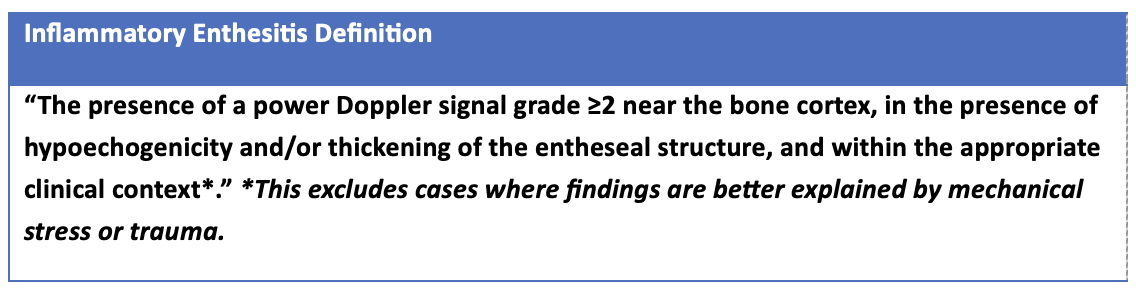 Table 1. New definition of inflammatory enthesitis at the single-enthesis level.
.jpg) Figure 1A . Number of elementary lesions identified in each category; 1B. Distribution of sonographic elementary lesions by category.
Figure 1A . Number of elementary lesions identified in each category; 1B. Distribution of sonographic elementary lesions by category.
.jpg) Figure 2. The prevalence of elementary lesions by enthesitis definition category. 1A. Hypoechogenicity (score 0-1); 1B. Enthesophyte (score 0-3); 1C.
Figure 2. The prevalence of elementary lesions by enthesitis definition category. 1A. Hypoechogenicity (score 0-1); 1B. Enthesophyte (score 0-3); 1C.
Thickening (score 0-1); 1D Calcification (Score 0-3); 1E. Erosion (score 0-1); 1F. Power Doppler (score 0-3). Chi-square test was performed to compare
the “definite enthesitis” and “uncertain” groups, with p-value < 0.05 marked with an asterisk (*) and a p-value < 0.01 marked with two asterisks.
To cite this abstract in AMA style:
Lucas Ribeiro A, Aydin S, Kaeley G, Afgani F, Bakewell C, Rosemffet M, Kohler M, Haddad A, Stoenoiu M, Polachek A, marin j, Katz A, Koppikar S, Eder L. Defining sonographic enthesitis in psoriatic arthritis: Developing a data- and expert-driven definition for inflammatory enthesitis at the single enthesis level [abstract]. Arthritis Rheumatol. 2025; 77 (suppl 9). https://acrabstracts.org/abstract/defining-sonographic-enthesitis-in-psoriatic-arthritis-developing-a-data-and-expert-driven-definition-for-inflammatory-enthesitis-at-the-single-enthesis-level/. Accessed .« Back to ACR Convergence 2025
ACR Meeting Abstracts - https://acrabstracts.org/abstract/defining-sonographic-enthesitis-in-psoriatic-arthritis-developing-a-data-and-expert-driven-definition-for-inflammatory-enthesitis-at-the-single-enthesis-level/