Session Information
Date: Sunday, October 26, 2025
Title: (0731–0764) Vasculitis – Non-ANCA-Associated & Related Disorders Poster I
Session Type: Poster Session A
Session Time: 10:30AM-12:30PM
Background/Purpose: Macrophages play a central role in the pathogenesis of large-vessel vasculitides, including giant cell arteritis (GCA) and Takayasu’s arteritis (TAK). Characterizing patterns of macrophage polarization induced by patient serum may uncover disease-specific immune signatures and therapeutic targets.
Methods: Purified monocytes from healthy donors were differentiated into macrophages (M0) using M-CSF and 10% fetal bovine serum for 4 days, followed by polarization toward M1 (IFNγ + LPS), M2a (IL-4 + IL-13), M2b (immune complexes + LPS), or M2c (IL-10 + dexamethasone) phenotypes. These in vitro-polarized macrophages served as the training dataset. Flow cytometry assessed CD40 (M1 marker), CD206 (M2a), HLA-DR (negative in M2b), and CD163 (M2c) expression. Macrophages exposed to serum from patients in remission with GCA (n=40), TAK (n=40), and healthy controls (HC, n=19) were analyzed (test samples). Data were extracted from FlowJo™ workspaces using CytoML and flowWorkspace in R and arcsinh-transformed. A K-Nearest Neighbors (KNN) classifier (k=10) trained on M0–M2c macrophages predicted phenotypes in test samples. Uniform Manifold Approximation and Projection (UMAP) was used for visualization, using 250 cells/sample for training and test samples, and all available cells for other analysis. Group-level differences were assessed using Fisher’s exact test, and per-sample differences with Mann–Whitney U tests.
Results: UMAP visualization revealed distinct clustering patterns among groups. Macrophages exposed to HC serum formed a relatively compact cluster, while those exposed to serum from patients with GCA and TAK showed greater dispersion, especially TAK, indicating broader phenotypic heterogeneity (Figure 1a-b). Spatial distribution of individual polarization markers is shown through UMAP overlays (Figure 1c-f).Quantitative analysis revealed that GCA samples retained the highest proportion of M0-like macrophages, followed by TAK, with HC showing the lowest. HC exhibited the highest proportions of M2a- and M2c-like macrophages, while TAK samples showed an increase in M2b-like macrophages. Despite tighter spatial clustering, HC macrophages polarized toward more varied phenotypes than the other groups (Figure 2). There were statistically significant differences among all groups, except for M1 between GCA and TAK. Although the proportion of M1 was negligible across all groups, HC showed a 6- and 4-fold increased prevalence compared to GCA and TAK, respectively.
Conclusion: Serum from patients with GCA and TAK induces distinct macrophage polarization profiles compared to HC. Serum in GCA maintains macrophages in an undifferentiated M0-like state, whereas serum in TAK promotes a modest M2b-skewed response associated with immune complex stimulation. In contrast, serum in HC predominantly favors regulatory and repair-associated macrophage phenotypes (M2a and M2c). These findings highlight macrophage phenotyping as a promising tool to uncover disease-specific immune dysregulation in large-vessel vasculitis.
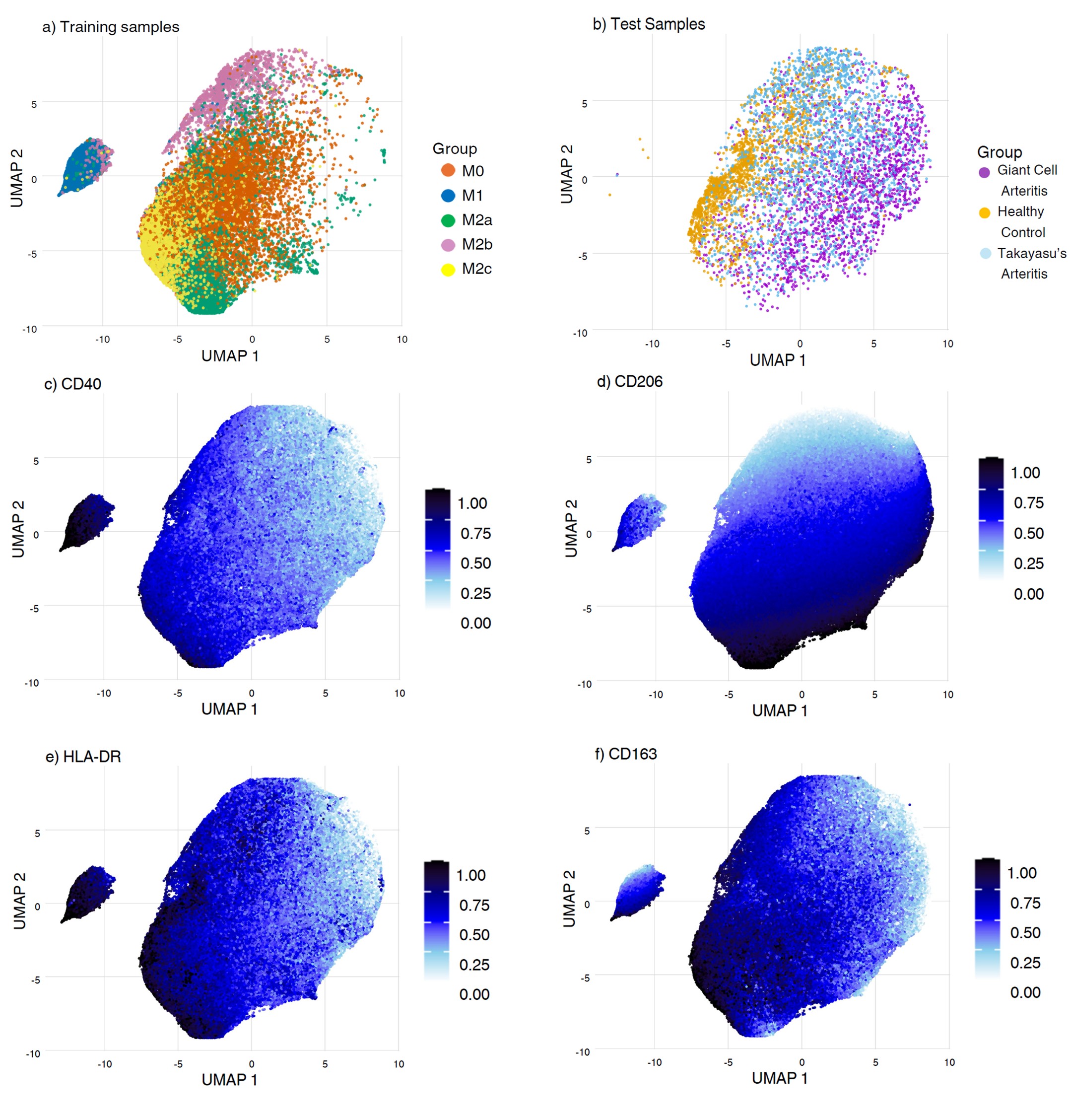 Figure 1: Uniform Manifold Approximation and Projection (UMAP) of training samples M0-M2c (a), test samples (b) and individual polarization markers (c-f).
.jpg) Figure 2: Percentage of each macrophage phenotype per group within all the cells of that group (a-c-e-g-i). Percentage of the median +/- interquartile range of each phenotype within each cell of the group (b-d-f-h-j). n.s. = non-significant; * = p-value < 0.05 – 0.01 ; ** = p-value < 0.01 – 0.001; *** = p-value < 0.001. GCA: giant cell arteritis; TAK: Takayasu’s arteritis; HC: healthy control.
Figure 2: Percentage of each macrophage phenotype per group within all the cells of that group (a-c-e-g-i). Percentage of the median +/- interquartile range of each phenotype within each cell of the group (b-d-f-h-j). n.s. = non-significant; * = p-value < 0.05 – 0.01 ; ** = p-value < 0.01 – 0.001; *** = p-value < 0.001. GCA: giant cell arteritis; TAK: Takayasu’s arteritis; HC: healthy control.
To cite this abstract in AMA style:
Carrión-Barberà I, Stultz R, Cuthbertson D, Khalidi N, Koening C, Langford C, McAlear C, Monach P, Moreland L, Pagnoux C, Seo P, Warrington K, Merkel P, Lood C. Patterns of Macrophage Polarization Induced by Serum from Patients with Giant Cell Arteritis and Takayasu’s Arteritis [abstract]. Arthritis Rheumatol. 2025; 77 (suppl 9). https://acrabstracts.org/abstract/patterns-of-macrophage-polarization-induced-by-serum-from-patients-with-giant-cell-arteritis-and-takayasus-arteritis/. Accessed .« Back to ACR Convergence 2025
ACR Meeting Abstracts - https://acrabstracts.org/abstract/patterns-of-macrophage-polarization-induced-by-serum-from-patients-with-giant-cell-arteritis-and-takayasus-arteritis/