Session Information
Session Type: Poster Session C
Session Time: 10:30AM-12:30PM
Background/Purpose: Imaging mass cytometry (IMC) is a powerful high throughput technique that enables in-situ multiparametric analysis of a single fixed tissue section while preserving spatial architecture. Sjögren’s Disease (SjD), despite being one of the most common systemic autoimmune disorders, has still unclear pathogenetic mechanisms involved and no effective treatment.
In the present study, we aim to perform in-situ deep immunophenotype at a spatially resolved cellular level of the inflammatory infiltrates of minor salivary gland (MSG) biopsies of SjD patients at different stages of histologic involvement and Sjögren related mucosa-associated lymphoid tissue (MALT) lymphomas.
Methods: To fulfil the purpose of the study, we collected 5 MSG biopsies of Sicca controls [(Focus Score (FS):0-1, Anti – Ro/SSA (-), Anti-La/SSB (-)], five MSG biopsies from SjD patients with mild infiltrates [FS:1-2, Tarpley Score (TS):1], five with intermediate (FS:2-4, TS:2) and five with severe (FS: 4-12, TS: 3-4). In addition, we also collected four MSG biopsies of patients with SjD associated MALT lymphomas. All demographic, clinical and serologic patient’s data were available. Formalin-fixed, paraffin-embedded (FFPE) tissue sections were prepared and stained using a panel of 27 metal conjugated antibodies. Serial FFPE tissue sections were used for the 2 panels. The acquisition process was performed using the Hyperion™ (Fluidigm) system. Hyperion data processing and analysis necessitated the use of several software in a labour-intensive pipeline. For the segmentation of cells, a validated proprietary artificial intelligence software was exploited (Youpi).
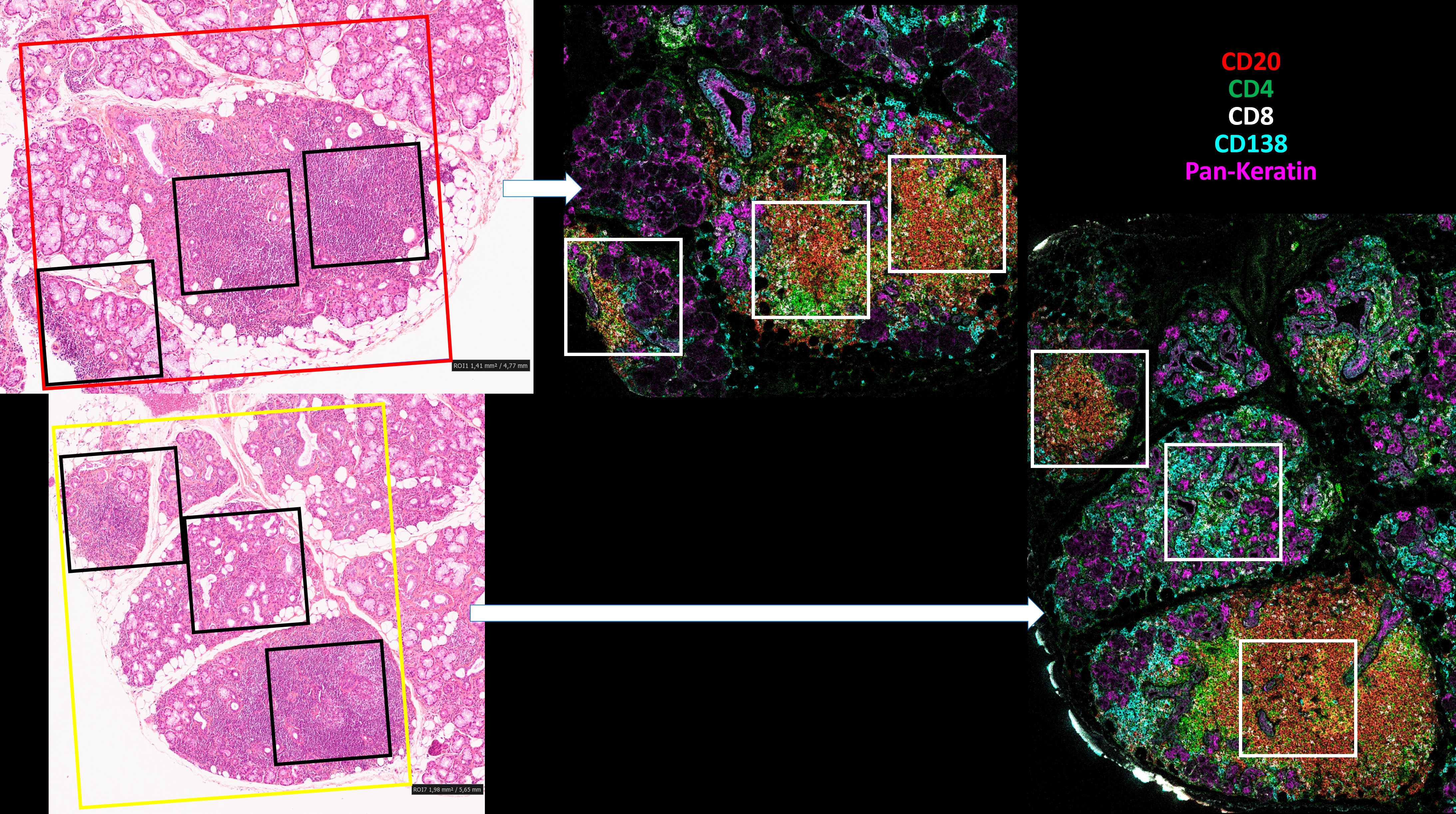
Results: One hundred and four areas of interest (ROIs) were analyzed. Each ROI represented an inflammatory infiltrate, and a new stratification of the ROIs was created based on the number of T and B cells. Unsupervised clustering revealed the presence of 21 immune cell populations. The cellular subpopulations of inflammatory infiltrates differed between different regions in the same patient (Figure 1). The presence of CD45RBhigh Double Negative B Cells (CD27– IgD–) and Podoplanin+ Double Negative B Cells appears to characterize the progression of disease severity towards lymphoma. Additionally, Double Negative B Cells and CD45RB+ Switched Memory B cells (CD27+ IgD–) were shown to occur almost exclusively in SjD-related lymphoma (Figure 2).
Conclusion: Advancing our knowledge on the target tissue, the variety of infiltrating cell subpopulations and their cell-to-cell interactions, previously limited by the low dimensionality of the histologic techniques available, might provide novel pathogenetic insights for the disease that can be targeted for the development of new treatment approaches for SjD. The use of IMC highlighted the role of Double Negative B cells which may play an important role in the pathogenesis of the disease and serve as potential biomarkers.
To cite this abstract in AMA style:
chatzis L, Hemon P, Scuiller Y, Goules A, Cornec D, Tzioufas A, Pers J. Deep Spatial Profiling Unveils Dominant Double Negative B Cells in Severe Forms of Sjögren’s Syndrome [abstract]. Arthritis Rheumatol. 2024; 76 (suppl 9). https://acrabstracts.org/abstract/deep-spatial-profiling-unveils-dominant-double-negative-b-cells-in-severe-forms-of-sjogrens-syndrome/. Accessed .« Back to ACR Convergence 2024
ACR Meeting Abstracts - https://acrabstracts.org/abstract/deep-spatial-profiling-unveils-dominant-double-negative-b-cells-in-severe-forms-of-sjogrens-syndrome/


