Session Information
Session Type: Poster Session C
Session Time: 10:30AM-12:30PM
Background/Purpose: Symptomatic knee osteoarthritis (OA) is more common in women than men. We sought to compare clinical and histological features and synovial gene expression between females and males with knee OA.
Methods: 135 patients undergoing Total Knee Arthroplasty (TKA) who met 1986 ACR classification criteria for knee OA were enrolled. Demographic and clinical characteristics including Erythrocyte Sedimentation Rate (ESR), high sensitivity C-Reactive Protein (hsCRP), Kellgren-Lawrence (KL) X-ray scores, and Osteoarthritis Research Society International (OARSI) cartilage scores were collected. Patient-reported pain was measured using the Knee Injury and Osteoarthritis Outcome Score (KOOS) pain subscale, Intermittent and Constant Osteoarthritis Pain Score (ICOAP), and pain Visual Analog Scale (VAS). Nine pathologist-scored synovial histology features were measured,1 and our computer vision pipeline was used to quantify cell density and cell aggregate number and size.2 RNA isolated from RNAlater-preserved synovium was used for bulk RNA-seq. cDNA libraries were prepared with KAPA-Stranded RNAseqKit with RiboErase, sequenced on a NovaSeq with 150 base pair paired end reads, targeting 100M reads per sample. Reads were pseudoaligned to Hg38 with Kallisto, and Limma was used to test for differentially expressed genes (DEGs). Gene set enrichment analysis (GSEA) and gene set variation analysis (GSVA) were used to test for differences in pathway and AMP phase 13 and 24 synovial cell subset enrichment according to sex.
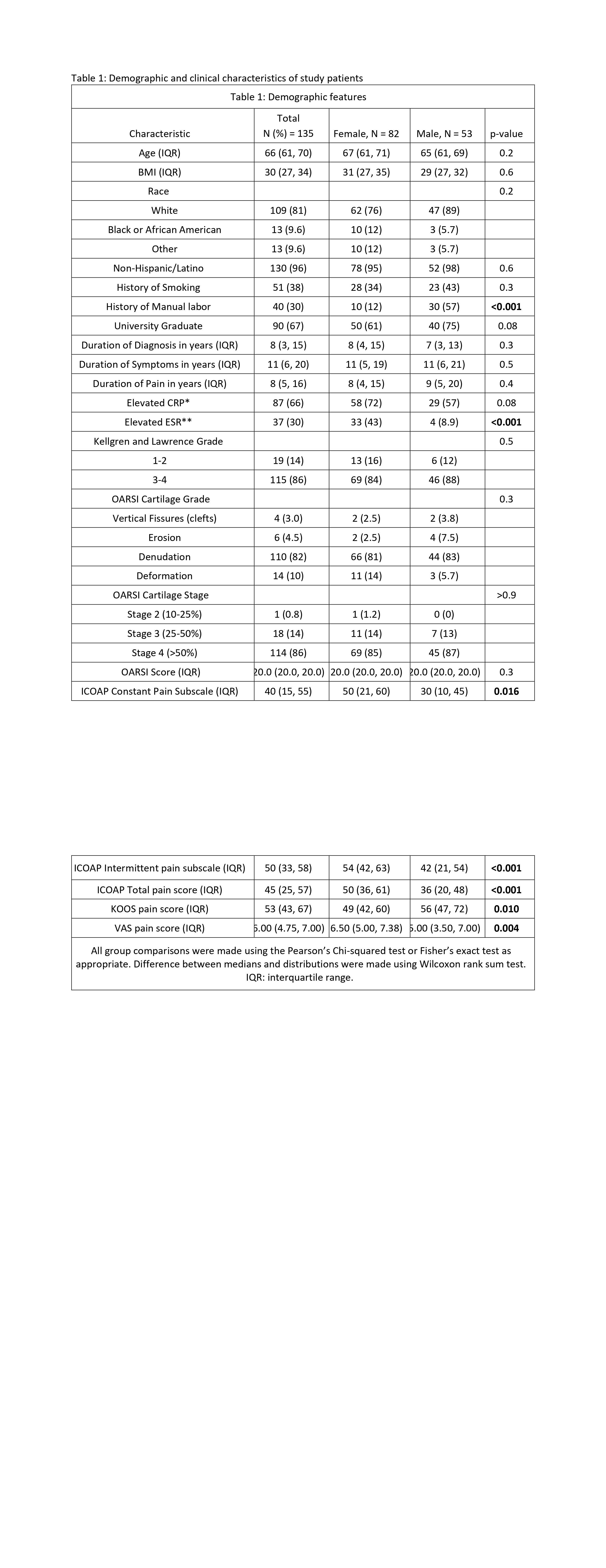
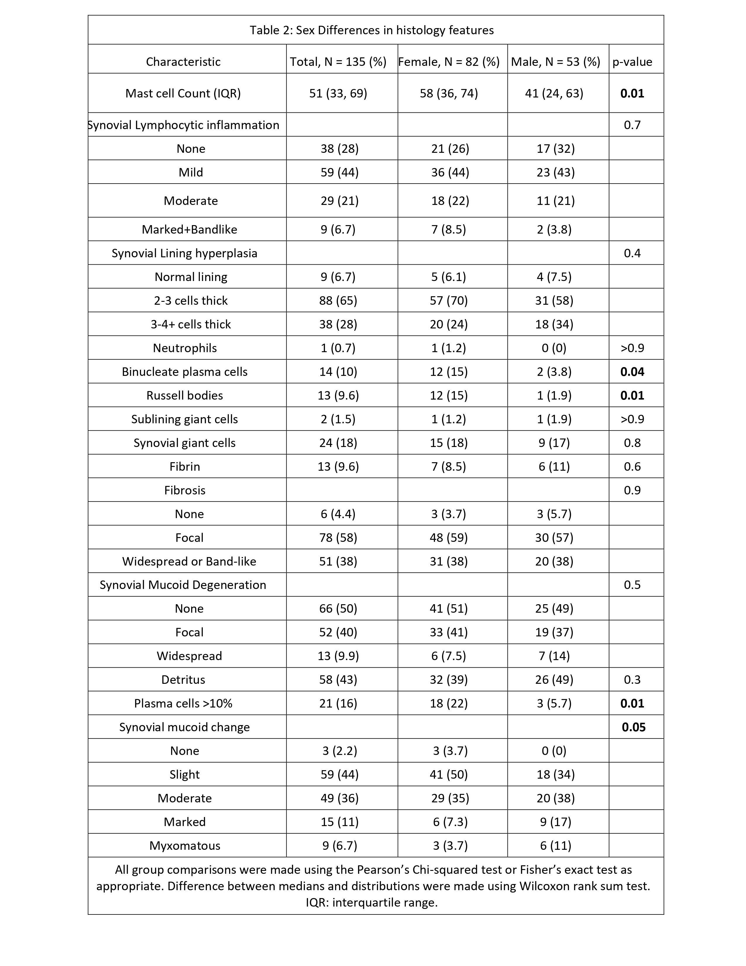
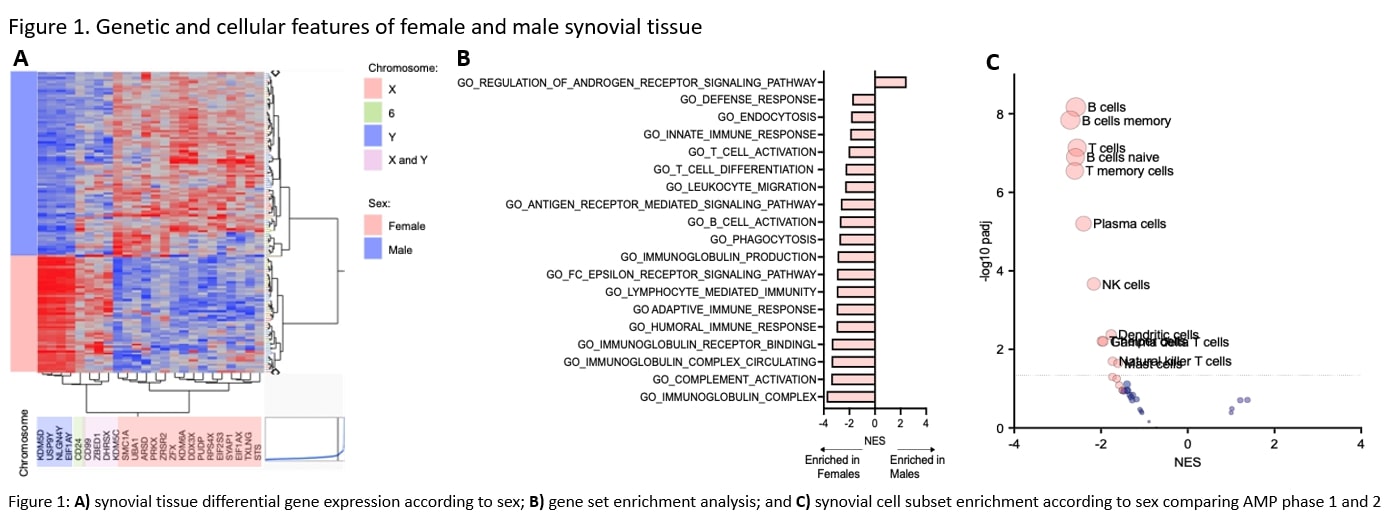
Results: A total of 61% (82) of enrolled subjects were female. While there were no significant differences in OA severity (KL grade), OARSI score, or symptom duration between sexes, females had increased ESR and reported worse pain (Table 1). Females had higher synovial plasma cell, binucleate plasma cell, and mast cell numbers, and increased scores of cell density, number, and inflammatory aggregates. Males had increased synovial mucoid degeneration (Table 2). RNA seq uncovered 25 DEGs between sexes, of which 24 were encoded on the X or Y chromosome, or both (Figure 1A). GSEA identified 132 pathways associated with inflammation and immune activation significantly enriched in females vs. males, including “adaptive immune response”, “complement activation”, “immunoglobulin complex” and “Fc epsilon receptor signaling pathways” (Figure 1B). One pathway involved in the regulation of androgen receptor signaling was significantly enriched in males. Synovial cell subset enrichment analyses showed increased B cell, T cell, NK cell, gd T cell, Nk T cell, Dendritic cells, and mast cell gene signatures in females (Figure 1C).
Conclusion: Despite similar OA severity and symptom duration, females had increased systemic and synovial tissue inflammation assessed by ESR, RNA-seq, and histology. Understanding the mechanisms underlying the association between sex, synovial inflammation and OA pain could provide therapeutic insight, and merits future investigation.
1) Orange et al, Arthritis Rheumatol 70, 690–701 (2018)
2) Frezza et al, Arthritis Rheumatol. 75, 2137–2147 (2023).
3&4) Zhang et al, Nat. Immunol. 20, 928–942 (2019) and Nature 623, 616–624 (2023).
To cite this abstract in AMA style:
Hale C, Otero M, Sun D, Parides M, Dicarlo E, Orange d, Wu Y, Robinson W, Mehta B, Cushing S, Sculco P, Maerz T, Malfait A, Ramirez D, Colbath A. Synovial Inflammation Is Increased in Females with Knee Osteoarthritis [abstract]. Arthritis Rheumatol. 2024; 76 (suppl 9). https://acrabstracts.org/abstract/synovial-inflammation-is-increased-in-females-with-knee-osteoarthritis/. Accessed .« Back to ACR Convergence 2024
ACR Meeting Abstracts - https://acrabstracts.org/abstract/synovial-inflammation-is-increased-in-females-with-knee-osteoarthritis/


