ACR Meeting Abstracts

ACR Meeting Abstracts

  • Meetings
    • ACR Convergence 2024
    • ACR Convergence 2023
    • 2023 ACR/ARP PRSYM
    • ACR Convergence 2022
    • ACR Convergence 2021
    • ACR Convergence 2020
    • 2020 ACR/ARP PRSYM
    • 2019 ACR/ARP Annual Meeting
    • 2018-2009 Meetings
    • Download Abstracts
  • Keyword Index
  • Advanced Search
  • Your Favorites
    • Favorites
    • Login
    • View and print all favorites
    • Clear all your favorites
  • ACR Meetings

Abstract Number: 1443

Natural Course of Degenerative Lesions of the Spine on Radiographs and MRI in Axial SpondyloarthritisFollowed 10 Years in the DESIR Cohort

Laura PINA VEGAS1, Miranda van Lunteren2, Damien Loeuille3, Caroline Morizot4, Esther Newsum5, Sofia Ramiro6, Floris van Gaalen7, Alain SARAUX8, Pascal Claudepierre9, Antoine Feydy10, Desiree van der Heijde11 and Monique Reijnierse1, 1Leiden University Medical Center, Leiden, Netherlands, 2Leiden University Medical Center, Leiden, Zuid-Holland, Netherlands, 3Phd, Nancy Vandoeuvre, Lorraine, France, 4Nancy University hospital, Nancy, France, 5Nij Smellinghe, Drachten, Netherlands, 6Leiden University Medical Center, Bunde, Netherlands, 7LUMC, Leiden, Zuid-Holland, Netherlands, 8CHU Brest, Brest, France, 9CHU Henri Mondor, AP-HP/EpiDermE, UPEC, Créteil, France, 10CHU Cochin, AP-HP, Paris, France, 11Department of Rheumatology, Leiden University Medical Center, Meerssen, Netherlands

Meeting: ACR Convergence 2024

Keywords: Epidemiology, Imaging, Magnetic resonance imaging (MRI), spondyloarthritis, X-ray

  • Tweet
  • Click to email a link to a friend (Opens in new window) Email
  • Click to print (Opens in new window) Print
Session Information

Date: Sunday, November 17, 2024

Title: SpA Including PsA – Diagnosis, Manifestations, & Outcomes Poster II

Session Type: Poster Session B

Session Time: 10:30AM-12:30PM

Background/Purpose: Radiographs and MRI are the two imaging techniques often used to assess structural lesions in axial spondyloarthritis (axSpA). A high prevalence of degenerative lesions (DLs) in the spine has been reported  among patients diagnosed with axSpA1 but their natural course is not well-known. Our aim was to investigate the DLs and their changes over 10 years (10Y) in patients with axSpA.

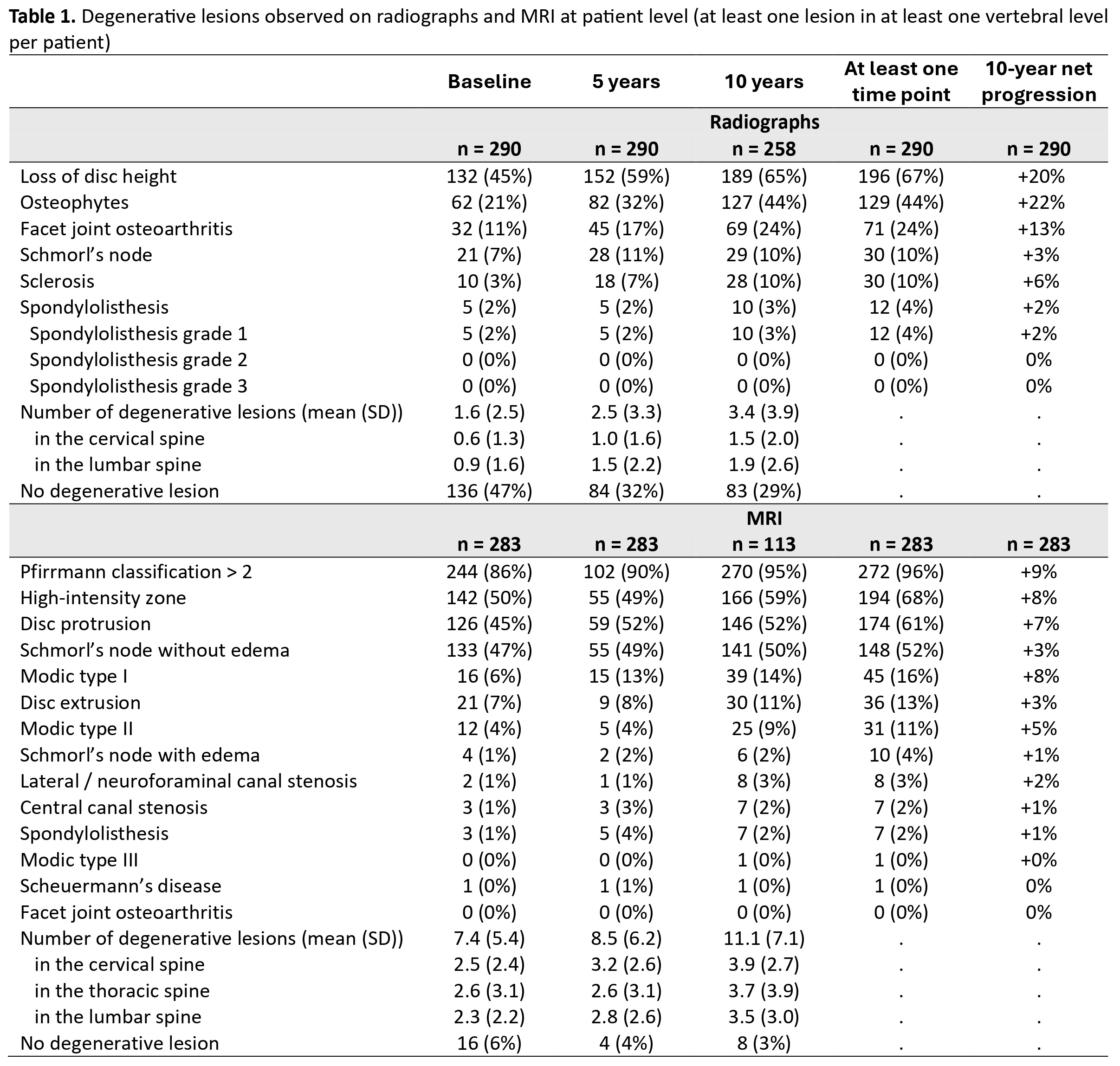
Methods: Cervical and lumbar spine radiographs and whole spine MRI at baseline, 5Y and 10Y of patients diagnosed with axSpA in the DESIR cohort were assessed for DLs by three central readers blinded to timepoint and to clinical, laboratory or any other imaging information1. Only patients with baseline and 10Y data were analyzed. The presence of each degenerative lesion was based on agreement between ≥2 out of 3 readers and an average of 3 central readers for continuous results was used (Table 1). Patient characteristics and the presence of DLs at the patient (≥1 lesion in ≥1 vertebral level) and at vertebral unit levels were reported. Net progression between 0 and 10Y was calculated by subtracting the patients that “improved” (with no specific lesion at 10Y in a patient who had ≥1 such lesion in ≥1 vertebral level at baseline) from those that “worsened” (with ≥1 specific lesion in ≥1 vertebral level at 10Y in a patient who had no such lesion at baseline) divided by the total number of patients. Finally, we compared the number of degenerative lesions at baseline and at 10Y on both imaging modalities using paired Student’s t-tests.

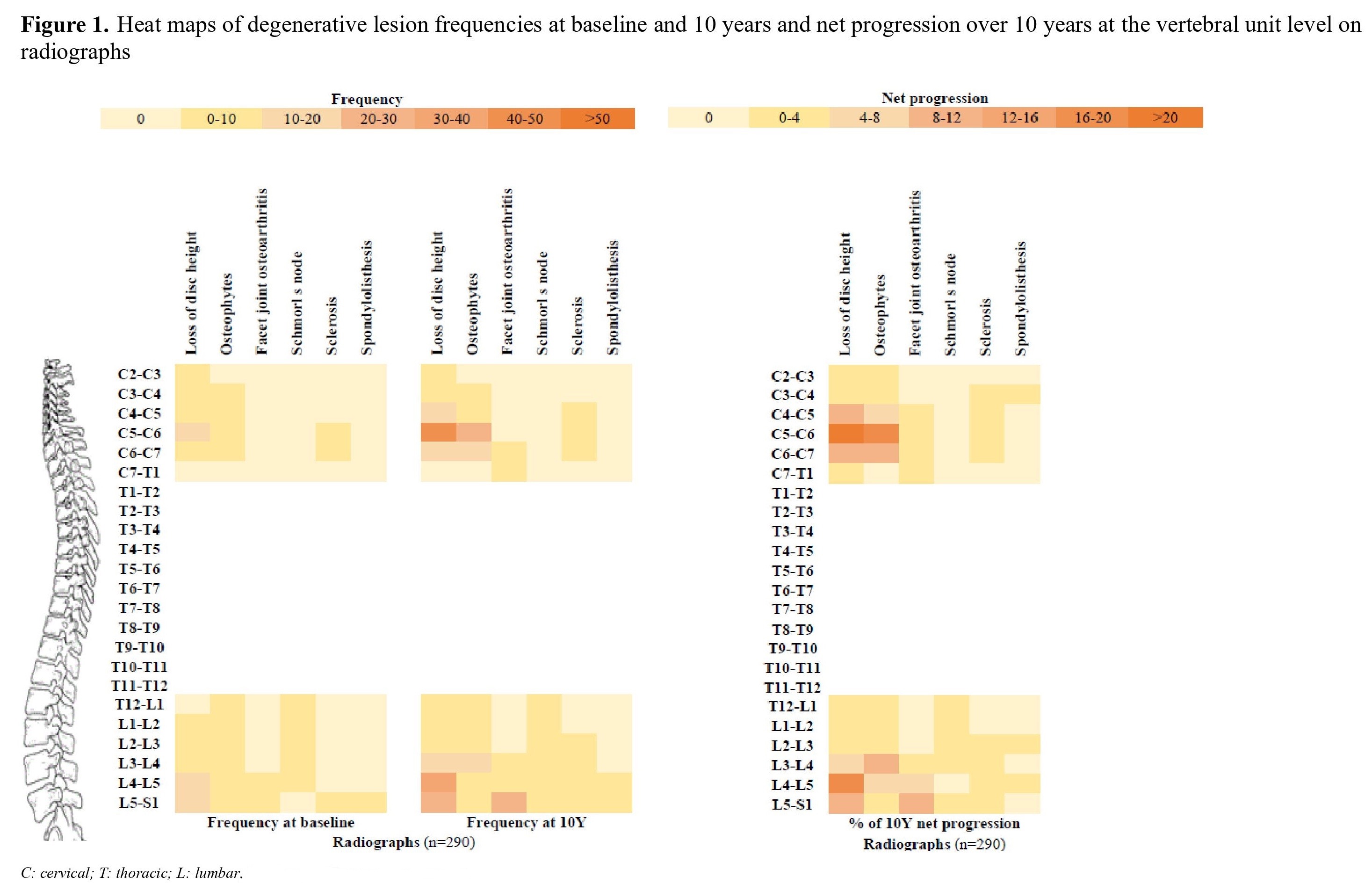
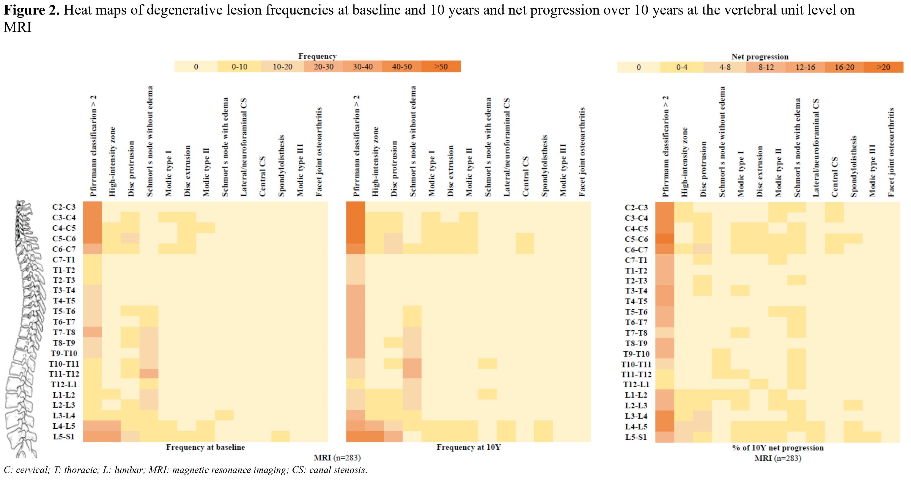
Results: Imaging was available for 291 patients (mean age [SD] 34.5 [8.6] years, 47% male). The most frequent DLs on radiographs were loss of disc height (45% at baseline, 65% at 10Y, net progression: +20%), followed by osteophytes (21% at baseline, 44% at 10Y, net progression: +22%) and facet joint osteoarthritis (11% at baseline, 24% at 10Y, net progression: +13%) (Table 1). An average of 1.6 (2.5) DLs per patient were observed on radiographs at baseline; this number increased to 3.4 (3.9) at 10Y (p-value < 0.0001). In total, 47% of patients had no degenerative lesions at baseline and 29% at 10Y. The most frequent degenerative lesions on MRI were disc degeneration (Pfirrmann classification >2: 86% at baseline, 95% at 10Y, net progression: +9%), followed by high-intensity zone (50% at baseline, 59% at 10Y, net progression: +8%), Schmorl’s node without edema (47% at baseline, 50% at 10Y, net progression: +3%) and disc protrusion (45% at baseline, 52% at 10Y, net progression: +7%) (Table 1). An average of 7.4 (5.4) DLs per patient were observed on MRI at baseline; this number increased to 11.1 (7.1) at 10Y (p-value < 0.0001). Only 6% of patients had no degenerative lesions at baseline and 3% at 10Y follow-up. The distribution of these spinal lesions is shown in the heat maps (Figures 1 and 2). 

Conclusion: The prevalence of spinal DLs is high in an inception cohort of axSpA with the total number of DLs increasing over 10Y in all parts of the spine on both radiographs and MRI. Therefore, in axSpA, DLs are important to be borne in mind when interpreting imaging, particularly after several years of follow-up.

Reference: 1. de Bruin F et al. RMD Open. 2018:4(1);e000657.

Supporting image 1

Loss of disc height was defined as narrowing of the disc space in comparison with two adjacent (healthy) discs. Osteophytes were described by reactive bone hypertrophy, seen as bony spurs arising from the vertebral body close to the vertebral endplate in a horizontal configuration (maximum of 45-degree angle with the endplate). Sclerosis was defined by an increased bone density and calcification adjacent to the vertebral endplates. Facet joint osteoarthritis was defined as sclerotic joint surfaces and osteophyte formation. Schmorl’s nodes were defined as a radiolucent contour defect of the vertebral endplate with sclerotic margins. Spondylolisthesis was defined as the slippage of one vertebral body with respect to the adjacent vertebral body. Disc degeneration was scored on a five-point scale (Pfirrmann classification), combining signal loss and loss of height of the intervertebral discs on STIR images. The high-intensity zone, indicating an annular tear or fissure, is seen as an area of high signal intensity located in the posterior annulus fibrosis on STIR images. Canal stenosis was defined as reduction of the anterior–posterior diameter of the spinal canal with compression on the spinal cord (at cervical and thoracic level) or as contact between the nerve root and disc material with obliteration of perineural intraforaminal fat or compression of the nerve root (at lumbar level). We considered bulging of a disc as either protrusion or extrusion; the distinction is based on whether the maximal diameter in any direction is at the base of the displacement (protrusion) or not (extrusion). Facet joint osteoarthritis was defined as sclerotic joint surfaces and osteophyte formation. Spondylolisthesis was defined as the slippage of one vertebral body with respect to the adjacent vertebral body. We used the Modic classification to assess degenerative lesions of the vertebral endplates. This is a three-point scale with type I defined as bone marrow oedema, type II is described as fatty changes and type III as sclerotic changes. Schmorl’s nodes were defined as an indentation of the (cranial or caudal) endplate with herniation of intervertebral disc material into the vertebra, with or without oedema. Scheuermann’s disease was defined as abnormal and excessive curvature of the spin with anterior wedging of greater than or equal to 5 degrees in 3 or more adjacent vertebral bodies. SD: standard deviation.

Supporting image 2

Supporting image 3


Disclosures: L. PINA VEGAS: Novartis, 5; M. van Lunteren: None; D. Loeuille: None; C. Morizot: None; E. Newsum: None; S. Ramiro: AbbVie, 1, 2, 5, 6, Alfasigma, 1, 2, 5, Galapagos, 1, 2, 5, Lilly, 1, 2, 6, MSD, 2, 5, 6, Novartis, 1, 2, 5, 6, Pfizer, 1, 2, 5, 6, UCB, 1, 2, 5, 6; F. van Gaalen: AbbVie, 12, Personal fees, BMS, 12, Personal fees, Eli Lilly, 12, Personal fees, Jacobus Stichting, 5, MSD, 12, Personal fees, Novartis, 2, 5, Stichting ASAS, 5, Stichting Vrienden van Sole Mio, 5, UCB Pharma, 5; A. SARAUX: Abbvie, BMS, Galapagos, Lilly, Novartis, Nordic, Pfizer, Roche-Chugai, Sanofi, UCB, 6, Abbvie, Bms, Lilly, Novartis, 5; P. Claudepierre: AbbVie, 2, 6, Amgen, 2, Biogen, 2, Celltrion, 2, Galapagos, 2, Janssen, 2, 6, Lilly, 2, 6, MSD, 2, 6, Novartis, 2, 6, Pfizer, 2, 6, UCB, 2; A. Feydy: None; D. van der Heijde: AbbVie, 2, ArgenX, 2, BMS, 2, Eli Lilly, 2, Galapagos, 2, GSK, 2, Imaging Rheumatology BV, 3, Janssen, 2, Novartis, 2, Pfizer, 2, Takeda, 2, UCB Pharma, 2; M. Reijnierse: ASAS, 2, ISS, 5.

To cite this abstract in AMA style:

PINA VEGAS L, van Lunteren M, Loeuille D, Morizot C, Newsum E, Ramiro S, van Gaalen F, SARAUX A, Claudepierre P, Feydy A, van der Heijde D, Reijnierse M. Natural Course of Degenerative Lesions of the Spine on Radiographs and MRI in Axial SpondyloarthritisFollowed 10 Years in the DESIR Cohort [abstract]. Arthritis Rheumatol. 2024; 76 (suppl 9). https://acrabstracts.org/abstract/natural-course-of-degenerative-lesions-of-the-spine-on-radiographs-and-mri-in-axial-spondyloarthritisfollowed-10-years-in-the-desir-cohort/. Accessed .
  • Tweet
  • Click to email a link to a friend (Opens in new window) Email
  • Click to print (Opens in new window) Print

« Back to ACR Convergence 2024

ACR Meeting Abstracts - https://acrabstracts.org/abstract/natural-course-of-degenerative-lesions-of-the-spine-on-radiographs-and-mri-in-axial-spondyloarthritisfollowed-10-years-in-the-desir-cohort/

Advanced Search

Your Favorites

You can save and print a list of your favorite abstracts during your browser session by clicking the “Favorite” button at the bottom of any abstract. View your favorites »

All abstracts accepted to ACR Convergence are under media embargo once the ACR has notified presenters of their abstract’s acceptance. They may be presented at other meetings or published as manuscripts after this time but should not be discussed in non-scholarly venues or outlets. The following embargo policies are strictly enforced by the ACR.

Accepted abstracts are made available to the public online in advance of the meeting and are published in a special online supplement of our scientific journal, Arthritis & Rheumatology. Information contained in those abstracts may not be released until the abstracts appear online. In an exception to the media embargo, academic institutions, private organizations, and companies with products whose value may be influenced by information contained in an abstract may issue a press release to coincide with the availability of an ACR abstract on the ACR website. However, the ACR continues to require that information that goes beyond that contained in the abstract (e.g., discussion of the abstract done as part of editorial news coverage) is under media embargo until 10:00 AM ET on November 14, 2024. Journalists with access to embargoed information cannot release articles or editorial news coverage before this time. Editorial news coverage is considered original articles/videos developed by employed journalists to report facts, commentary, and subject matter expert quotes in a narrative form using a variety of sources (e.g., research, announcements, press releases, events, etc.).

Violation of this policy may result in the abstract being withdrawn from the meeting and other measures deemed appropriate. Authors are responsible for notifying colleagues, institutions, communications firms, and all other stakeholders related to the development or promotion of the abstract about this policy. If you have questions about the ACR abstract embargo policy, please contact ACR abstracts staff at [email protected].

Wiley

  • Online Journal
  • Privacy Policy
  • Permissions Policies
  • Cookie Preferences

© Copyright 2025 American College of Rheumatology