Session Information
Date: Sunday, November 17, 2024
Title: Metabolic & Crystal Arthropathies – Basic & Clinical Science Poster II
Session Type: Poster Session B
Session Time: 10:30AM-12:30PM
Background/Purpose: In gout, monosodium urate (MSU) crystal deposition occurs preferentially at certain joints, most frequently the first metatarsophalangeal (MTP) joint. Dual energy CT (DECT) allows visualization of MSU crystals. The aim of this DECT study was to map the distribution of MSU crystal deposition within the MTP joints in people with tophaceous gout. The study hypothesis was that MSU crystal deposition is evenly distributed within the joint in tophaceous gout.
Methods: Bilateral feet DECT scans of 124 people with tophaceous gout (57% male, mean disease duration 21 years, 98% on urate-lowering therapy, mean serum urate 6.9mg/dL) were analyzed. All participants met the ACR/EULAR 2015 classification criteria for gout. The metatarsal head and phalangeal base of each MTP joint was analyzed by dividing each bone into four quadrants (dorsal, plantar, medial, and lateral) as described by Pecherstorfer et al (ACR Open Rheumatology 2020). Each bone quadrant was scored for the presence or absence of MSU crystal deposition (eight sites per joint, 80 sites per participant). MSU crystal deposition was considered present if in contact with or directly adjacent (within 1mm) to bone or cartilage in at least two planes. All images were analyzed independently by two trained readers (inter-reader kappa 0.78 and percentage agreement 98.9%), and differences between readers was resolved by image review. Data was analyzed using general estimating equations undertaken to adjust for the likely closer agreement between bones in the same individual.
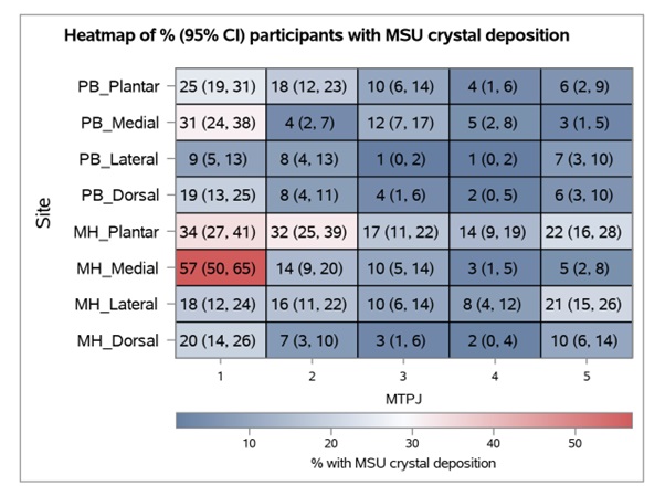
Results: There was marked variation in MSU crystal deposition across the analyzed sites (Figure). The medial quadrant of the 1st metatarsal head was most affected (57%) and significantly more affected than any other analyzed site. The next most common affected sites were the plantar quadrants of the 1st and 2nd metatarsal heads and medial quadrant of the 1st phalangeal base (all >30%). MSU crystal deposition was very rare in the phalangeal base of the 3rd and 4th MTP joints, particularly the lateral quadrants (1%). Across all joints, there was more MSU crystal deposition at the 1st MTP joint compared to other joints, at the metatarsal head compared to the phalangeal base, and at the plantar and medial quadrants compared to other quadrants (P< 0.001 for all comparisons).
Conclusion: In people with tophaceous gout, MSU crystal deposition is not evenly distributed. There is marked lack of uniformity with preferential sites of deposition not only between different joints, but also within joints. Several biological mechanisms may explain these findings, including the influence of localized tissue planes, temperature or microtrauma.
To cite this abstract in AMA style:
De Silva C, Díaz C, Gamble G, Horne A, Doyle A, Stamp L, Dalbeth N. Mapping Monosodium Urate Crystal Deposition Within Joints in Tophaceous Gout: A Dual Energy CT Study [abstract]. Arthritis Rheumatol. 2024; 76 (suppl 9). https://acrabstracts.org/abstract/mapping-monosodium-urate-crystal-deposition-within-joints-in-tophaceous-gout-a-dual-energy-ct-study/. Accessed .« Back to ACR Convergence 2024
ACR Meeting Abstracts - https://acrabstracts.org/abstract/mapping-monosodium-urate-crystal-deposition-within-joints-in-tophaceous-gout-a-dual-energy-ct-study/

