Session Information
Session Type: Poster Session B
Session Time: 10:30AM-12:30PM
Background/Purpose: Sjögren’s Disease (SjD) is a systemic autoimmune disease that primarily targets salivary and lacrimal glands, causing tissue deterioration and loss of function. Disease progression depends on a plethora of scarcely explored cellular interactions. Over time, infiltrating immune cells organize into tertiary lymphoid structures (TLS) bearing self-reactive B cells. In some patients, this immune dysregulation ultimately leads to B cell lymphoma. However, the gene expression changes correlating with disease progression remain largely unknown. We aimed to elucidate the alterations in gene expression pathways and cellular heterogeneity in salivary glands of patients with SjD over the course of the disease, uncovering their association with disease phenotypes.
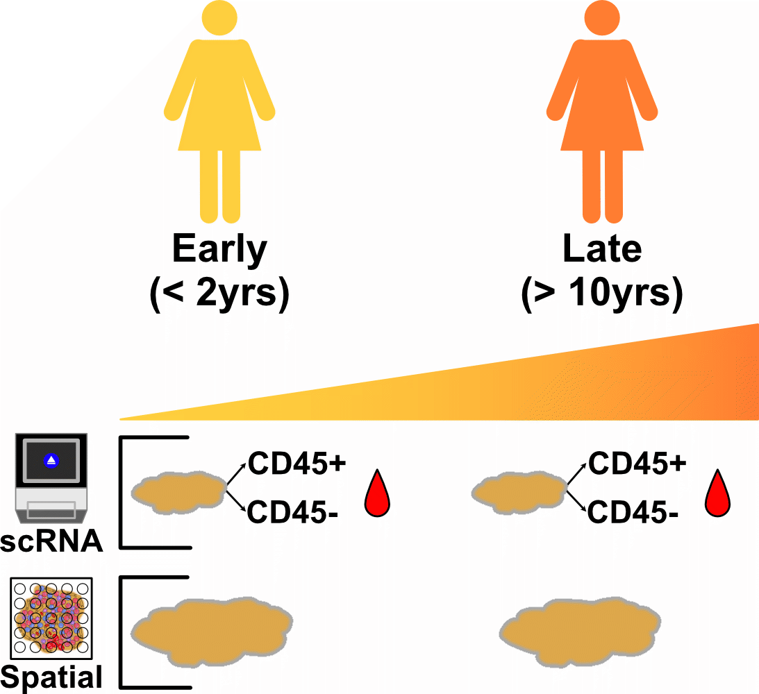
Methods: We recruited SjD patients at early (< 2 years since diagnosis) and late ( >10 years) disease stages. Minor salivary gland biopsies were collected from these patients. From those biopsies, we performed single-cell transcriptomics (scRNA-seq) and spatial transcriptomics (ST) (Fig. 1). Following quantification, scRNA-seq and ST data were analyzed using the Seurat R package.
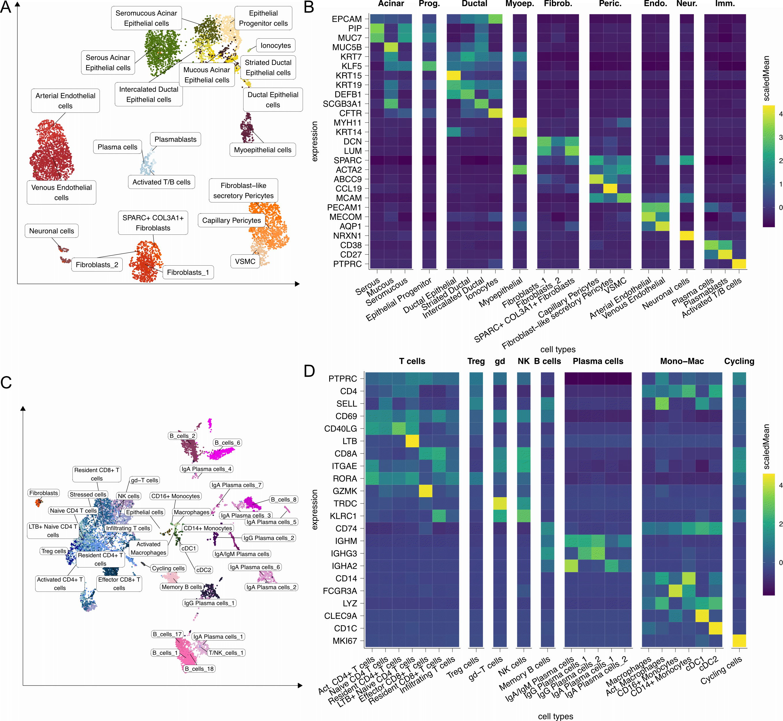
Results: Analysis of scRNA-seq data from non-hematopoietic cells (sorted as CD45-) showed the cell types characteristic of normal human salivary glands (Fig.2 A and B). The hematopoietic cells sorted (as CD45+) from the same biopsies also showed a wide variety of cell types, with an abundance of plasma cells and various T cell populations (Fig.2 C and D).
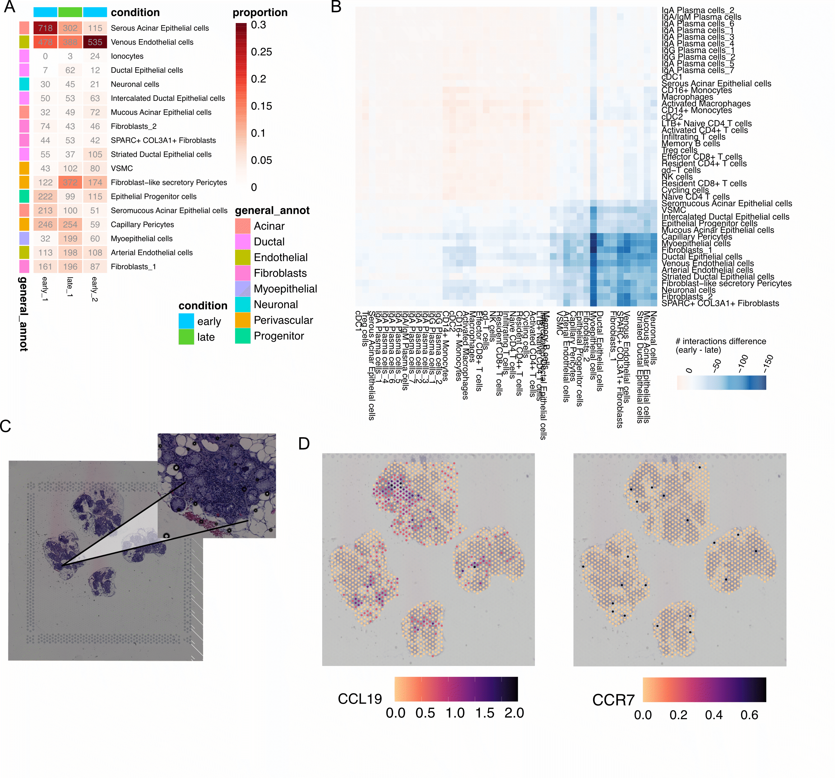
Salivary gland cell types were mostly consistent between early and late donors. Fibroblast-like secretory pericytes were among those enriched in a late disease stage (Fig. 3A), and involved in more predicted cell-cell interactions compared to early donors (Fig. 3B). Salivary gland ST data from a late donor, showed that CCL19 (a chemoattractant ligand for CCR7, present in naïve T cells), was specifically expressed by fibroblast-like secretory pericytes (Fig. 2B), and present in and around areas of immune cell infiltration (Fig.3 C and D).
Conclusion: We generated a cell and tissue transcriptomic atlas of salivary glands from SjD patients at different disease stages. Our analyses revealed a wide range of cell types, including a fibroblast-like secretory pericyte population, and suggest that alterations to its abundance may have a pathogenical role in disease progression. The increased number of predicted cell-cell interactions and association with immune cell infiltrates further suggest a role for these cells in immune cell recruitment and TLS organization.
To cite this abstract in AMA style:
Gomes T, Bandeira M, do Amaral Vieira R, Correia B, Barreira S, Gaspar P, Cruz-Machado R, Filipe B, Ávila-Ribeiro P, Ribeiro F, Vidal B, Val B, Costa F, Teodósio Chícharo A, Silva A, Silvério-Antonio M, Forjaz Lacerda J, Eurico Fonseca J, López Presa D, Khmelinskii N, Kumar S, Graca L, C. Romão V. Unraveling the Progression of Sjögren’s Disease at the Cellular and Histological Level Using Single-cell and Spatial Transcriptomics [abstract]. Arthritis Rheumatol. 2024; 76 (suppl 9). https://acrabstracts.org/abstract/unraveling-the-progression-of-sjogrens-disease-at-the-cellular-and-histological-level-using-single-cell-and-spatial-transcriptomics/. Accessed .« Back to ACR Convergence 2024
ACR Meeting Abstracts - https://acrabstracts.org/abstract/unraveling-the-progression-of-sjogrens-disease-at-the-cellular-and-histological-level-using-single-cell-and-spatial-transcriptomics/


