Background/Purpose: Rheumatoid arthritis (RA) is synonymous for involving synovial lined structures such as joints and tendon sheaths. Alterations in the extensor tendons overlying the metacarpophalangeal (MCP) joints which do not have synovial sheaths and which do not normally communicate with the joint recess have been observed with sonography but have not been clearly characterized. We report findings from a sonographic pilot study regarding changes in the MCP extensor tendons in patients with RA. These findings will lead to further study of the etiology and importance of these findings.
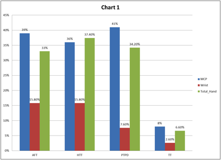
Methods: A retrospective cross-sectional study was done on 26 RA patients who had ultrasounds (US) of their hands between June 2010 and April 2012. Scans of the dorsal wrist and dorsal and volar 2nd- 5th MCP joints had been recorded in these patients. Esaote MyLab 70 ultrasound machine was used with a 12-18MHz LA 435 probe. The studies were re-read by GSK and B-mode and Power Doppler (PD) joint activity were scored. Changes in the extensor tendons were examined for the presence of B-Mode ultrastructural changes and PD. B- mode changes included anechoic fluid around the tendons (AFT), hypoechoic tissue around the tendon (HTT), tendon thickening (TT), tendon rupture (TR) and tendon calcifications (TC). The presence of vascularity around the tendon- Peritendon power doppler (PTPD) was recorded dichotomously. Joint erosions and tenosynovitis was also recorded.
Results: 26 patients, 77% were women, mean age of 56 years, mean body mass index of 31 and mean duration of disease 159 months were studied. PTPD and AFT were the highest findings, 41% and 39% respectively, observed around the extensor tendons at the level of the MCP. At the wrist, distal to the retinaculum, AFT and HHT were the dominant findings in 15.8 % of hands. TT was the least common finding, being found in 8% of hands at the MCP and 2.6% at the wrist. Joint erosions were noted in 50% of hands at the wrist. One patient had PD signal around the annular hood of the extensor tendon complex. B-Mode and Power Doppler were scored semiquantitatively from 0-3. Levels of activity were stratified in the following tertiles, 0-0.9, 1-1.9 and 2-3.The overall level of disease activity was high. Forty- four percent of patients had B-Mode score (BMS) in the upper tertile at the MCPs and 88% had a BMS in the upper tertile at the wrist. Twenty- eight percent of the patients had a Power Doppler score (PDS) in the upper tertile at the MCPs, and 67% at the wrist. Although the greatest joint activity was noted at the wrist, the extensor tendons at the MCPs displayed the greatest frequency of pathological changes.
Conclusion: Significant changes at the dorsal extensor tendon of the MCP joints were AFT, HTT, PTPD and TT. Although these findings have been reported in the past, they have not been fully characterized. Further studies are needed to define the etiology and significance of these findings.
Disclosure:
L. A. Ramrattan,
None;
G. S. Kaeley,
None.
« Back to 2013 ACR/ARHP Annual Meeting
ACR Meeting Abstracts - https://acrabstracts.org/abstract/ultrasound-characteristics-of-extensor-tendon-abnormalities-and-peritendinous-fluid-in-rheumatoid-arthritis/

