ACR Meeting Abstracts

ACR Meeting Abstracts

  • Meetings
    • ACR Convergence 2024
    • ACR Convergence 2023
    • 2023 ACR/ARP PRSYM
    • ACR Convergence 2022
    • ACR Convergence 2021
    • ACR Convergence 2020
    • 2020 ACR/ARP PRSYM
    • 2019 ACR/ARP Annual Meeting
    • 2018-2009 Meetings
    • Download Abstracts
  • Keyword Index
  • Advanced Search
  • Your Favorites
    • Favorites
    • Login
    • View and print all favorites
    • Clear all your favorites
  • ACR Meetings

Abstract Number: 1035

Diagnostic and Educational Utility of an Ultrasound Protocol for Evaluation of Hand Pain in Systemic Sclerosis

Meridith Balbach, Tracy Frech, Nicholson Chadwick, Kevin Byram and Erin Chew, Vanderbilt University Medical Center, Nashville, TN

Meeting: ACR Convergence 2023

Keywords: education, patient, hand, Musculoskeletal Examination, Systemic sclerosis, Ultrasound

  • Tweet
  • Click to email a link to a friend (Opens in new window) Email
  • Click to print (Opens in new window) Print
Session Information

Date: Monday, November 13, 2023

Title: (1033–1051) Imaging of Rheumatic Diseases Poster I

Session Type: Poster Session B

Session Time: 9:00AM-11:00AM

Background/Purpose: Hand impairment is a prevalent issue among systemic sclerosis (SSc) patients and contributes to disability and diminished quality of life. Managing hand pain can be challenging due to the coexistence of various manifestations such as inflammatory arthritis, tendinopathies, joint contractures, sclerodactyly, calcinosis, acro-osteolysis, Raynaud’s phenomenon (RP), digital ulcers (DU), and chronic pain. The physical examination and radiographs are the primary methods for evaluating hand pain but are limited in scope. We hypothesized that joint ultrasound (US) is more sensitive than combined joint exam and hand radiograph for identification of acro-osteolysis, articular disease (synovitis, erosions, osteophytes), and periarticular disease (tendinopathies, calcinosis) and can assist in targeted treatment strategies.

Methods: Five randomly selected SSc patients with nonspecific hand pain were referred for musculoskeletal US exam. SSc clinical features were recorded. Joint US was compared to physical exam features of synovitis, tendinopathy, digital ulcers, or calcinosis and radiographic features of erosions, osteophytes, and acro-osteolysis. A pre- and post-ultrasound survey assessed patient and provider perception of diagnostic understanding and treatment needs.

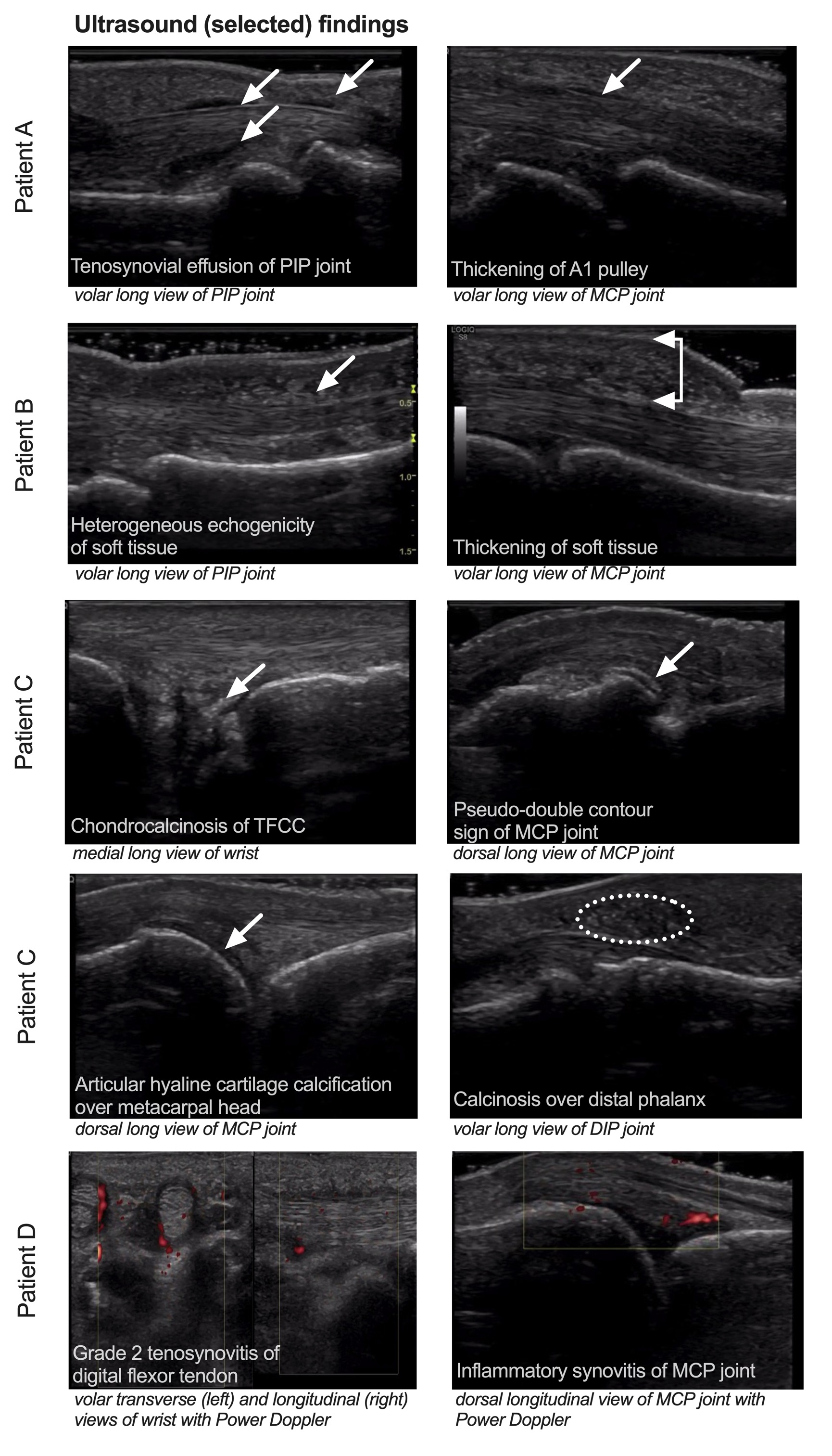
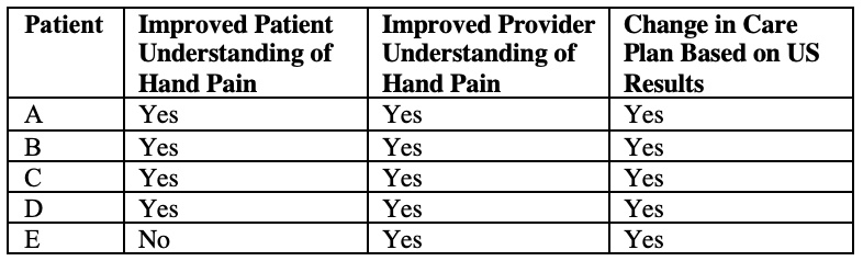
Results: Baseline characteristics, exam findings, radiographs, and ultrasound findings are noted in Table 1. No patients had digital ulcers or calcinosis on physical exam or acro-osteolysis on radiograph. Ultrasound exam was able to detect diverse manifestations of hand pathology (Figure 1). Ultrasound review improved the referring provider’s confidence in treatment decision improved in all cases (Table 2). Patient A was diagnosed with mechanical osteoarthritis, patient B with sclerodactyly, patient C with calcium pyrophosphate arthropathy, Patient D with inflammatory arthritis, and Patient E with chronic pain. The treatment plan was modified in all five patients. US improved patients’ understanding of the cause of their joint pain in four patients. After undergoing US, all patients reported feeling that their joint problem had been more thoroughly examined and being more likely to adhere to their provider’s treatment recommendations.

Conclusion: The physical exam and radiographs are not sufficient for the evaluation of hand pain in systemic sclerosis. Standardized US assessment in SSc can accurately evaluate hand pain and quantify acro-osteolysis, articular disease, and periarticular disease. Detection of these features can improve provider confidence in management decisions and advance patient understanding of disease, potentially improving treatment adherence and outcomes.

Supporting image 1

Table 1: Patient clinical characteristics and physical exam, radiograph, and ultrasound findings.
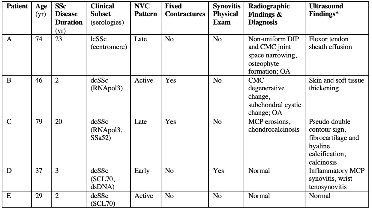
Yr= years; lcSSc= limited cutaneous systemic sclerosis; dcSSc= diffuse cutaneous systemic sclerosis; NVC= nailfold videocapillaroscopic pattern; DIP= distal interphalangeal; CMC= carpometacarpal; OA= osteoarthritis; MCP= metacarpophalangeal; CPPD= calcium pyrophosphate deposition.
*Summarized ultrasound findings are displayed in Table 1 with specific pathologic features detected on ultrasound represented in Figure 1.
PIP= proximal interphalangeal; MCP= metacarpophalangeal; TFCC= triangular fibrocartilage complex; DIP= distal interphalangeal

Supporting image 2

Figure 1: Selected pathologic findings on musculoskeletal ultrasound exam for patients A-D. Relevant features are indicated by white arrows or dotted oval and imaging view is labeled in italics.
PIP= proximal interphalangeal; MCP= metacarpophalangeal; TFCC= triangular fibrocartilage complex; DIP= distal interphalangeal

Supporting image 3

Table 2: Patient and provider perception survey results after ultrasound exam.


Disclosures: M. Balbach: None; T. Frech: None; N. Chadwick: None; K. Byram: None; E. Chew: None.

To cite this abstract in AMA style:

Balbach M, Frech T, Chadwick N, Byram K, Chew E. Diagnostic and Educational Utility of an Ultrasound Protocol for Evaluation of Hand Pain in Systemic Sclerosis [abstract]. Arthritis Rheumatol. 2023; 75 (suppl 9). https://acrabstracts.org/abstract/diagnostic-and-educational-utility-of-an-ultrasound-protocol-for-evaluation-of-hand-pain-in-systemic-sclerosis/. Accessed .
  • Tweet
  • Click to email a link to a friend (Opens in new window) Email
  • Click to print (Opens in new window) Print

« Back to ACR Convergence 2023

ACR Meeting Abstracts - https://acrabstracts.org/abstract/diagnostic-and-educational-utility-of-an-ultrasound-protocol-for-evaluation-of-hand-pain-in-systemic-sclerosis/

Advanced Search

Your Favorites

You can save and print a list of your favorite abstracts during your browser session by clicking the “Favorite” button at the bottom of any abstract. View your favorites »

All abstracts accepted to ACR Convergence are under media embargo once the ACR has notified presenters of their abstract’s acceptance. They may be presented at other meetings or published as manuscripts after this time but should not be discussed in non-scholarly venues or outlets. The following embargo policies are strictly enforced by the ACR.

Accepted abstracts are made available to the public online in advance of the meeting and are published in a special online supplement of our scientific journal, Arthritis & Rheumatology. Information contained in those abstracts may not be released until the abstracts appear online. In an exception to the media embargo, academic institutions, private organizations, and companies with products whose value may be influenced by information contained in an abstract may issue a press release to coincide with the availability of an ACR abstract on the ACR website. However, the ACR continues to require that information that goes beyond that contained in the abstract (e.g., discussion of the abstract done as part of editorial news coverage) is under media embargo until 10:00 AM ET on November 14, 2024. Journalists with access to embargoed information cannot release articles or editorial news coverage before this time. Editorial news coverage is considered original articles/videos developed by employed journalists to report facts, commentary, and subject matter expert quotes in a narrative form using a variety of sources (e.g., research, announcements, press releases, events, etc.).

Violation of this policy may result in the abstract being withdrawn from the meeting and other measures deemed appropriate. Authors are responsible for notifying colleagues, institutions, communications firms, and all other stakeholders related to the development or promotion of the abstract about this policy. If you have questions about the ACR abstract embargo policy, please contact ACR abstracts staff at [email protected].

Wiley

  • Online Journal
  • Privacy Policy
  • Permissions Policies
  • Cookie Preferences

© Copyright 2025 American College of Rheumatology