Session Information
Date: Monday, November 13, 2023
Title: (0934–0964) Systemic Sclerosis & Related Disorders – Basic Science Poster
Session Type: Poster Session B
Session Time: 9:00AM-11:00AM
Background/Purpose: Systemic Sclerosis (SSc) is an autoimmune disease characterized by vascular and immune system dysfunction, along with tissue fibrosis. Vascular damage has been recognized as the pathological basis of SSc, but there are no medicines approved by FDA for SSc early vasculopathy treatment for the underlying mechanism remains elusive. Our previous study found GRB2 was downregulated by salvianolic acid B, a small molecule drug that attenuated skin fibrosis of SSc by protecting endothelial cells from oxidative stress injury. Here we aim to investigate the role of GRB2 in the vasculopathy of SSc.
Methods: The microarray data of SSc skin biopsies in Caucasians were obtained from the Gene Expression Omnibus (GEO) database (GSE95065). The expression of GRB2 was further detected in Chinese SSc patients (n = 20) and healthy controls (n = 20). To further explore the possible effects of Grb2 on skin fibrosis in vivo, Grb2 siRNA was used to lower the expression of Grb2 in the BLM-induced skin fibrosis mouse model. The apoptosis of EA.hy926 endothelial cells was induced by H2O2 and apoptosis ratio was measured by flow cytometric. Transcriptome and phosphoproteomic analyses in EA.hy926 cells transfected with NC and GRB2 siRNA were performed to explore the GRB2 regulated pathway.
Results: The expression of GRB2 was significantly enhanced in SSc patient skin, 1.51-fold in Caucasians and 1.40-fold in Chinese. The immunohistochemical staining showed that there were more GRB2 positive cells in SSc patients than in normal controls, and double immunofluorescence staining showed the endothelial cells of SSc patient’s skin highly expressed GRB2 (Figure 1). Then the in vivo study revealed that in situ apoptosis of endothelium increased under the treatment of BLM, while interfering Grb2 could reduce endothelial cell apoptosis, also proved by the protein expression of cleaved caspase-3 (Figure 2). The in vitro study showed that decreased GRB2 expression level could up-regulate tight junction related genes expression and inhibit apoptosis to protect endothelial cells from H2O2-induced hyperpermeability (Figure 3). Moreover, both transcriptome and phosphoproteomic analysis suggested the focal adhesion pathway was enriched in GRB2 siRNA transfected endothelial cells (Figure 3).
Conclusion: Our results demonstrated GRB2 is highly expressed in endothelial cells of SSc skin, and inhibiting GRB2 could effectively attenuate endothelial cell apoptosis and BLM-induced skin fibrosis. GRB2 may regulate focal adhesion and VEGF signaling pathways to induce apoptosis of endothelial cells. GRB2 is expected to be a new therapeutic target for SSc.
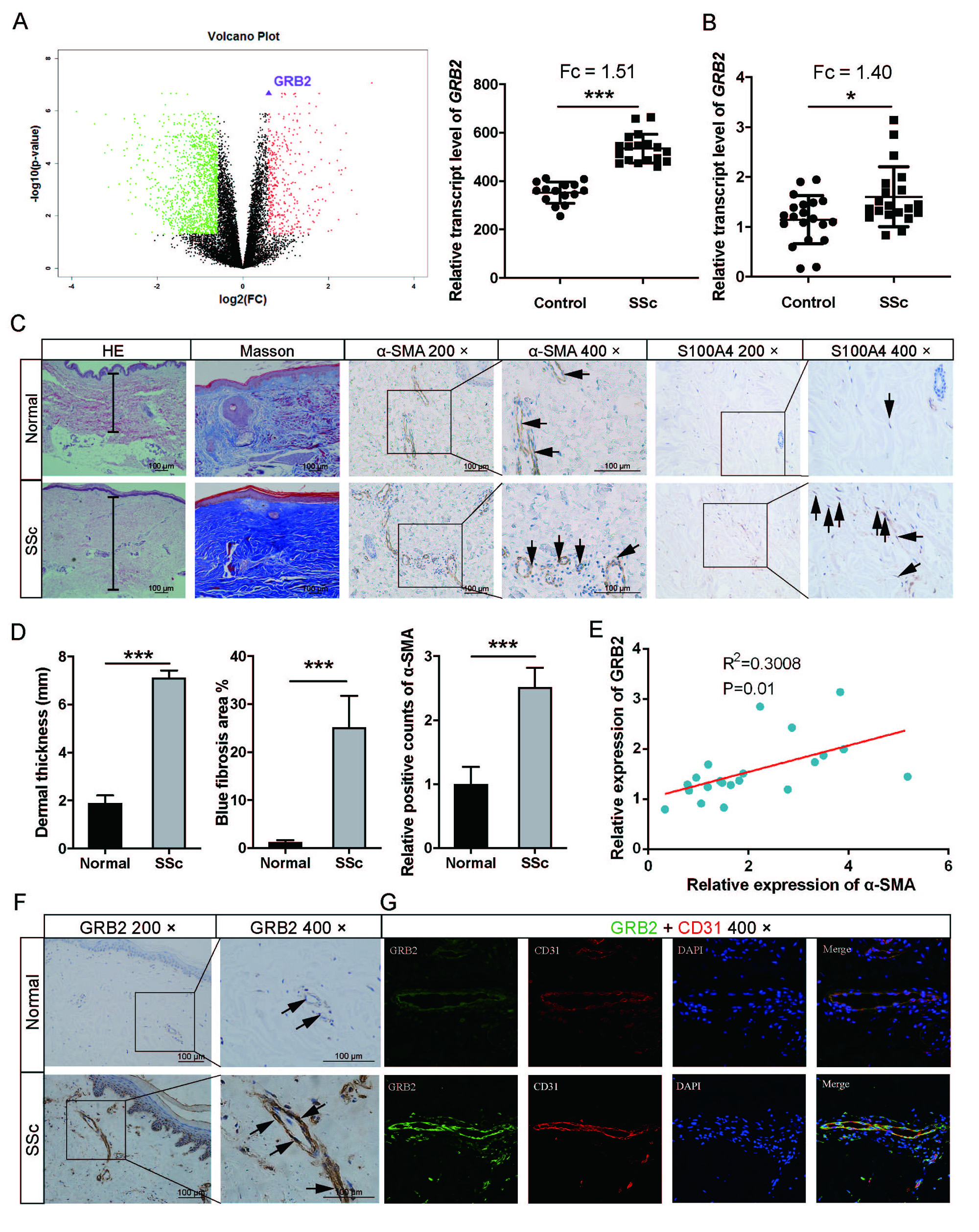
(A) Left: The volcano plot of differential expression genes between normal and SSc. Right: Relative transcription levels of GAB1 in the skin lesions from the GEO database. (B) Relative transcription levels of GAB1 in the Chinese skin lesions. (C) HE, Masson’s staining, and IHC staining for healthy control and SSc patients. Black arrows indicate α-SMA/S100A4 positive staining cells. (D) The left plot shows dermal thickness calculated by measuring the distance between the dermal-epidermal junction and the dermal–subcutaneous fat junction. The middle plot shows the blue fibrosis area of Masson’s staining for healthy control and SSc patients. The right plot shows the relative positive counts of α-SMA of IHC staining for healthy control and SSc patients. (E) The association of GRB2 and α-SMA expression levels were analyzed. (F) IHC staining for GRB2 in the skin of healthy control and SSc patients. Black arrows indicate GRB2 positive staining cells. (G) GRB2 and CD31 in the skin were labeled by double immunofluorescence staining, and the cell nucleus was stained with DAPI. Data are presented as mean ± SD in two groups and compared by t-test. *P < 0.05, *** P < 0.001.
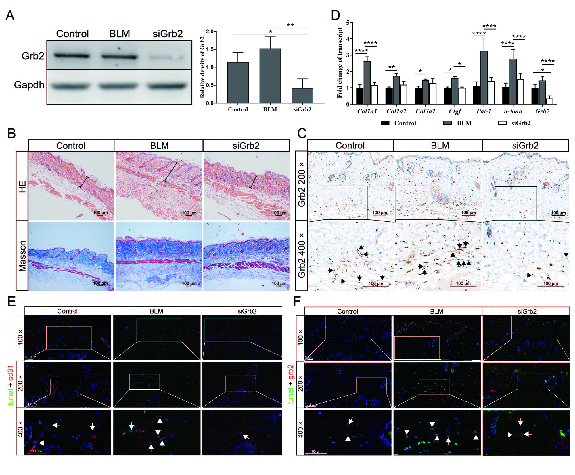
(A) Grb2 protein expression level in the dorsal skin of mice was determined by western blot analysis and analyzed by ImageJ. (B) IHC staining for Grb2 in the mouse skin of different groups. The black arrows indicate Grb2-positive staining cells. (C) IHC staining of the dorsal skin of mice in different groups showed the relative positive number of Grb2. (D) Relative transcription levels of Grb2 and fibrotic genes in mouse skin were measured by real-time RT-PCR. (E) Double immunofluorescence staining of tunel and cd31 in normal, BLM, and siGrb2 groups. The white arrows indicate tunel-cd31-positive staining cells. (F) Double immunofluorescence staining of tunel and grb2 in different groups. Arrows indicate the positive cells. Data are presented as mean ± SD of three samples and compared with one-way ANOVA; *P < 0.05, ** P < 0.01, *** P < 0.001.
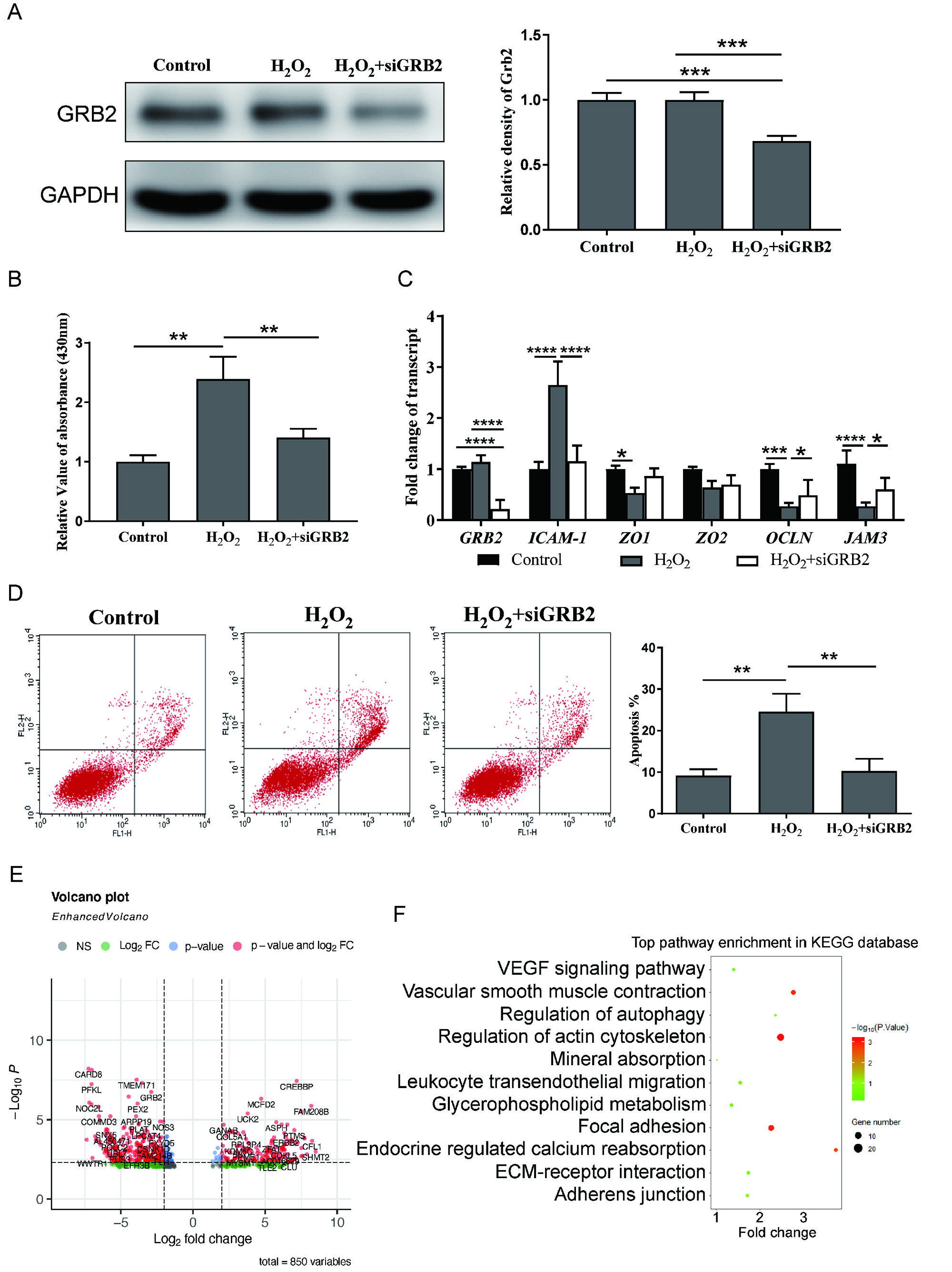
(A) The protein level of GRB2 was determined by western blotting and quantified by ImageJ. (B) Effects of siGRB2 on H2O2-induced endothelial permeability in EA.hy926 cells detected by cell permeability assay. (C) The relative mRNA levels of ICAM1, ZO1, ZO2, OCLN, and JAM3 were detected by real-time RT-PCR in different groups. (D) The ratio of apoptosis detected by flow cytometry in the cells of the control, H2O2, and H2O2 + siGRB2 groups. The column plot shows the apoptosis ratio in the different groups. (E) The Volcano plot of differentially expressed genes (DEGs) in NC or GRB2 siRNA transfected EA.hy926 cells. (F) KEGG pathway analysis of DEGs. Data are presented as means ± SD of three samples and compared with one-way ANOVA; *P < 0.05, ** P < 0.01, *** P < 0.001.
To cite this abstract in AMA style:
Huang Y, Zhao H, Shi X, Liu J, Lin J, Ma Q, Jiang S, Pu W, Ma Y, Liu J, Wu W, Wang J, Liu Q. GRB2 Serves as a Viable Target Against Skin Fibrosis in Systemic Sclerosis by Regulating Endothelial Cell Apoptosis [abstract]. Arthritis Rheumatol. 2023; 75 (suppl 9). https://acrabstracts.org/abstract/grb2-serves-as-a-viable-target-against-skin-fibrosis-in-systemic-sclerosis-by-regulating-endothelial-cell-apoptosis/. Accessed .« Back to ACR Convergence 2023
ACR Meeting Abstracts - https://acrabstracts.org/abstract/grb2-serves-as-a-viable-target-against-skin-fibrosis-in-systemic-sclerosis-by-regulating-endothelial-cell-apoptosis/