ACR Meeting Abstracts

ACR Meeting Abstracts

  • Meetings
    • ACR Convergence 2024
    • ACR Convergence 2023
    • 2023 ACR/ARP PRSYM
    • ACR Convergence 2022
    • ACR Convergence 2021
    • ACR Convergence 2020
    • 2020 ACR/ARP PRSYM
    • 2019 ACR/ARP Annual Meeting
    • 2018-2009 Meetings
    • Download Abstracts
  • Keyword Index
  • Advanced Search
  • Your Favorites
    • Favorites
    • Login
    • View and print all favorites
    • Clear all your favorites
  • ACR Meetings

Abstract Number: 2179

Spatial and Single-Cell Transcriptomics Identify Alcam+ Macrophage / CD6+ T-Cell Interactions and Accumulation of IgG2b+ Class-Switched Plasma Cells in Marco+ Medullary Sinuses of Joint-Draining Popliteal Lymph Nodes in TNF-Tg Mice with Advanced Arthritis

H. Mark Kenney1, Yue Peng2, Kiana Chen2, Javier Rangel-Moreno2, Elizabeth Pritchett2, Jeffrey Fox2, Benjamin Korman3, Jennifer Anolik2, Lianping Xing4, Christopher Ritchlin5, Edward Schwarz2 and Chia-Lung Wu2, 1University of Rochester Medical Center, Henrietta, NY, 2University of Rochester Medical Center, Rochester, NY, 3University of Rochester, Rochester, NY, 4University of Rochester Medical Center, Webster, NY, 5Allergy, Immunology and Rheumatology Division, University of Rochester Medical School, Canandaigua, NY

Meeting: ACR Convergence 2022

Keywords: Bioinformatics, Mouse Models, RA, rheumatoid arthritis

  • Tweet
  • Click to email a link to a friend (Opens in new window) Email
  • Click to print (Opens in new window) Print
Session Information

Date: Monday, November 14, 2022

Title: Abstracts: RA – Animal Models

Session Type: Abstract Session

Session Time: 3:00PM-4:00PM

Background/Purpose: Severe inflammatory-erosive arthritis in tumor necrosis factor transgenic (TNF-Tg) mice is associated with B-cell translocation into sinuses of joint-draining popliteal lymph nodes (PLNs) via unknown mechanisms (1). Thus, we characterized transcriptional changes in PLNs of TNF-Tg mice with Early vs. Advanced stages of arthritis combining spatial and single-cell transcriptomics to assess the potential mechanisms orchestrating B-cell translocation.

Methods: PLNs from TNF-Tg male mice with Early (5-7-months, n=6 PLNs) and Advanced (Adv, >8-months, n=12 PLNs) arthritis were processed for spatial transcriptomics. In addition, 8-9k cells from pooled PLNs of Early and Adv TNF-Tg groups were subjected to droplet-based single-cell RNA-sequencing (scRNAseq, n=6 PLNs/group) and immune profiling scRNAseq (n=2 PLNs/group, 2 replicates). Data were analyzed with Seurat, NicheNet (cell interaction), and scRepertoire (clonality) R packages. PLN immunofluorescence (IF) and ankle μCT measures were utilized to validate RNA detection and evaluate bone loss, respectively.

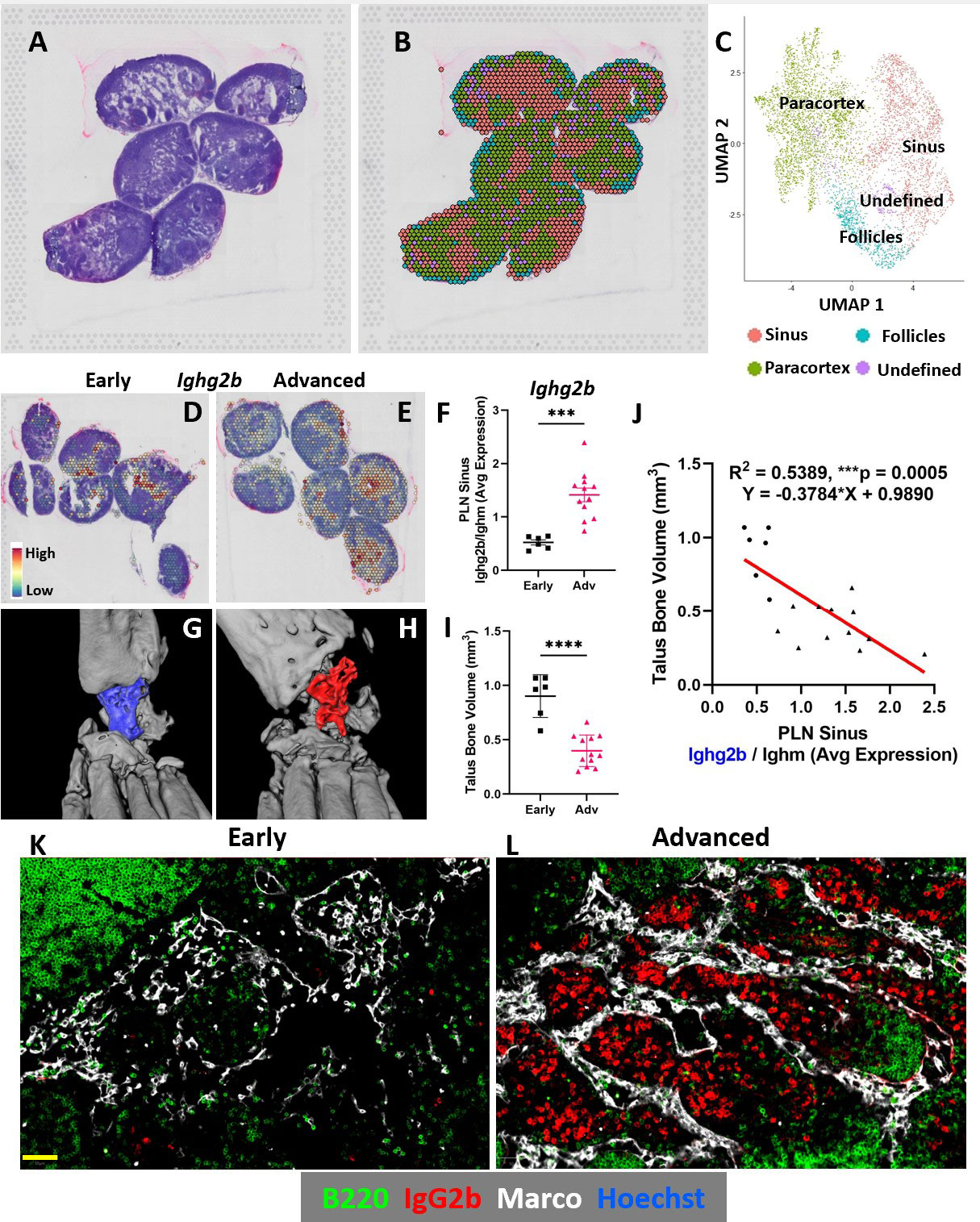
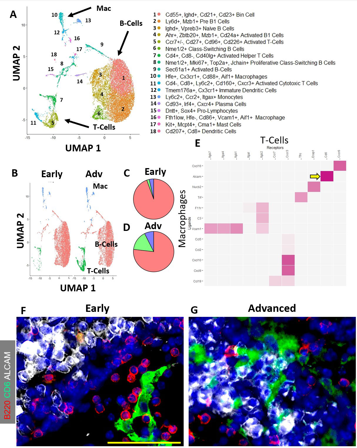
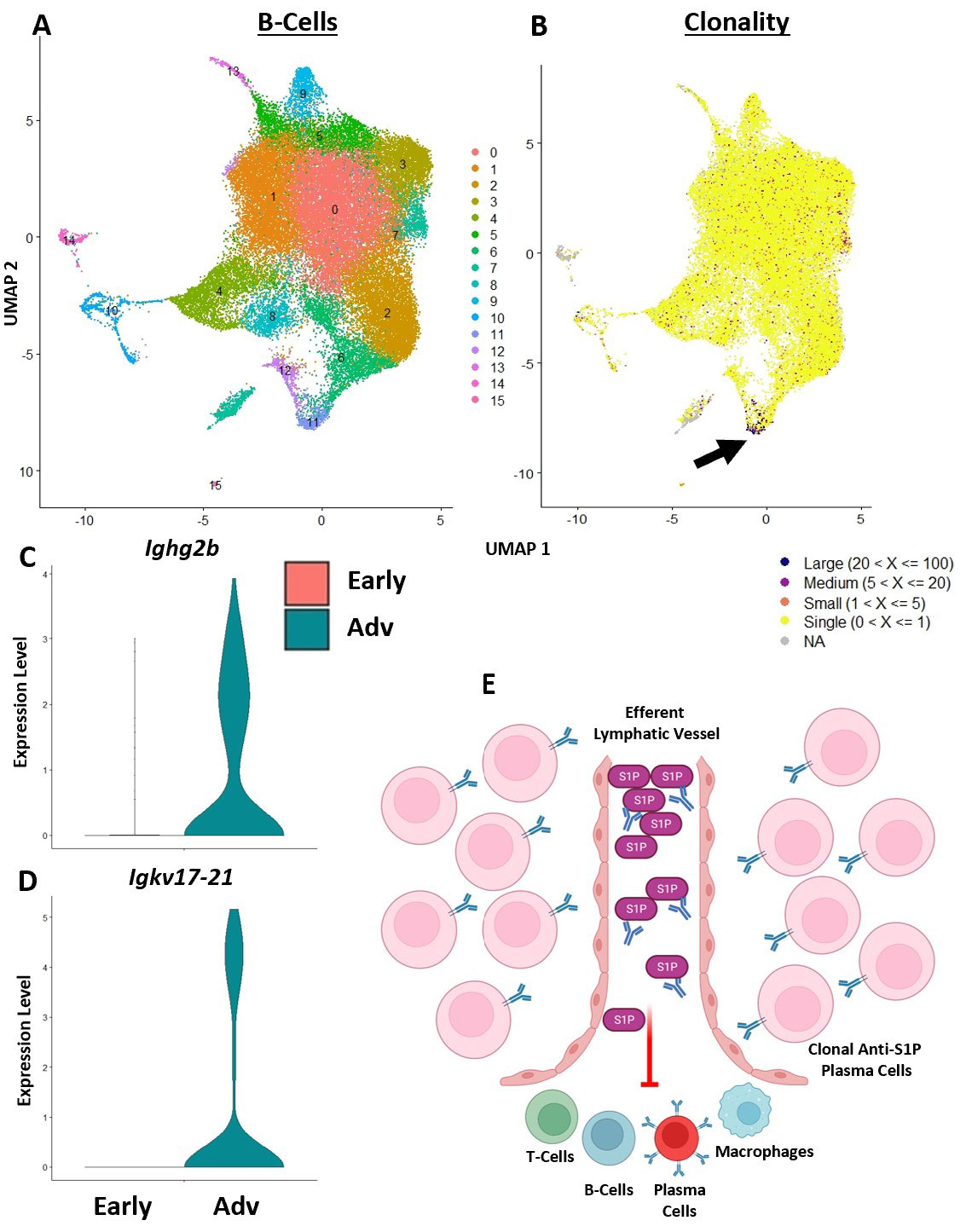
Results: Spatial transcriptomics revealed a significant and selective increase in Ighg2b/Ighm expression ratio in PLN sinuses of Adv vs Early conditions (0.5±0.1 vs 1.4±0.5 counts/counts; p< 0.001). Ighg2b expression significantly correlated with increased bone erosions and reduced talus bone volumes in the afferent ankle joint (R2=0.54, p< 0.001). IF confirmed the accumulation of IgG2b+/B220– plasma cells adjacent to Marco+ peri-follicular medullary sinuses (Figure 1). scRNAseq resolved 18 distinct cell populations with a dramatic increase in T-cells in Adv vs Early PLNs (15.8% vs 2.0%). Bioinformatics indicated a high probability interaction between Alcam+ macrophages and CD6+ T-cells. IF confirmed the close proximity of Alcam+/CD6+ cells only in the Adv condition (Figure 2). Assessment of B-cell clonality revealed 15 polyclonal clusters and a single oligoclonal population ( >20 cells of same clonotype). Differential gene expression analysis of the oligoclonal population showed increased Ighg2b expression along with unique variable light chain expression (Igkv17-21) in the Adv condition, which has been previously associated with antigen specificity to sphingosine-1-phosphate (S1P) (2) (Figure 3).

Conclusion: B-cell translocation into PLN sinuses is associated with the generation of IgG2b+ plasma cells in Advanced but not Early arthritis. These B-cell changes are concomitant with T-cell accumulation and activation via Alcam+/CD6+ interaction. Bioinformatic analysis and previous studies (2) suggest the presence of clonal S1P-specific plasma cells that may compromise S1P-dependent cell egress in PLNs and alter lymphatic function in inflammatory arthritis. Ongoing studies will confirm the existence of S1P-specific plasma cells and evaluate the therapeutic potential of CD6 blockade in TNF-Tg arthritis, similar to recent clinical trials (3).

References

1. Bouta et al. Nat Rev Rheumatol. 14(2):94-106. 2018.
2. Farokhi et al. Antibodies (Basel). 9(2):10. 2020.
3. Rodriguez et al., Clin Exp Immunology. 191(2):229-39. 2018.

Supporting image 1

Figure 1. Increased IgG2b expression in PLN sinuses correlates with reduced bone volume in the afferent ankle joint. PLNs from TNF-Tg mice were harvested and processed for spatial transcriptomics. A representative H&E-stained image (A) corresponds to a transcriptional representation (B) of the PLNs from an Advanced capture area where the sinus regions are shown by red spots (C). Differential gene expression of Ighg2b (IgG2b) within the sinus regions are shown by a spatial feature plot overlying the H&E-stained PLNs from Early (D) and Advanced (E) groups with a significant increase in Ighg2b/Ighm expression in Advanced sinuses (p<0.001) (F). Ex-vivo micro-CT was performed on the hindpaws of TNF-Tg mice with Early (G) and Advanced (H) arthritis with a significant reduction in talus bone volumes in Advanced TNF-Tg mice (p<0.0001) (I). Linear regression analysis demonstrated negative correlation between PLN sinus Ighg2b expression and talus bone volumes in the afferent ankles (R2 = 0.54, p<0.001) (J). The increase in IgG2b+ cells in Advanced vs Early TNF-Tg PLNs was validated by immunofluorescence. IgG2b+ cells are limited in Early PLNs (K), while Advanced arthritis is associated with a dramatic accumulation of IgG2b+ (red) / B220- (green) plasma cells localized to the Marco+ (white) PLN peri-follicular medullary sinuses (L). Statistics: Unpaired t-test (F, I); linear regression (J); ***p<0.001, ****p<0.0001. Scale bar (yellow) = 50μm (K, L).

Supporting image 2

Figure 2. Increased IgG2b+ plasma cells in Advanced PLNs with predicted Alcam+ macrophage and CD6+ T-cell co-stimulation. Single-cell suspensions were collected from Early and Advanced TNF-Tg PLNs (n=3 mice, 6 PLNs pooled/group), flow sorted for Hoechst+ / Sytox- live cells, and processed for scRNAseq. Following integration of the Early (8192 cells) and Advanced (8136 cells) samples, unsupervised clustering in Seurat resolved 18 distinct cell clusters (A), representing subtypes of B-cells, T-cells, and macrophages (B). Comparison of Early vs Advanced PLNs demonstrated a substantial increase in the number of T-cells (green; Early 2.0% (C) vs Adv 15.8% (D)). A remarkable increase was also noted in the proportion of macrophages in Advanced PLNs (3.6% Early (C) vs 7.4% Advanced (D)). Note the color of the pie chart matching the color of unique cell clusters in the UMAP shown in B. To elucidate potential T-cell / macrophage co-stimulatory pathways in Advanced PLNs, cell interaction analysis (NicheNet / Seurat) was performed, which indicated high interaction potential between Alcam (macrophages, ligand) and CD6 (T-cells, receptor) (E, yellow arrow). PLN immunohistochemistry further demonstrated a notable physical separation between Alcam+ and CD6+ cells in the Early condition (F), while these cells were found interacting in close proximity within Advanced PLNs (G). Scale bar (yellow) = 50μm (F, G).

Supporting image 3

Figure 3. Clonal B-cells in Advanced PLNs express BCRs with known anti-S1P specificity and potential to inhibit immune cell egress. Immune profiling scRNAseq was performed on Early and Advanced PLNs (n=2 experiments/group, n=2 PLNs pooled, n=8-9k cells/experiment) following cell sorting for Hoechst+ / Sytox- live cells. Cells from all scRNAseq experiments were integrated and the B-cells were subset for reclustering, which resolved 16 distinct cell populations (A). The B-cell receptors (BCRs) were analyzed (scRepertoire / Seurat), and a clonal population of B-cells (B, black arrow, dark blue spots, >20 B-cells with the same clonotype) localized to cluster 11 in A. Differential gene expression analysis of the oligoclonal cluster 11 demonstrated Ighg2b class-switching (C) with unique light-chain variable region gene expression of Igkv17_21 in the Advanced condition (D), which has been previously suggested to promote antigen specificity for sphingosine_1 phosphate (S1P) (2). Thus, a subset of the IgG2b+ B-cells accumulated in the PLN medullary region and associated with arthritic severity (Figure 1) may exhibit anti-S1P specificity that inhibits immune cell egress through joint-draining lymph nodes (E). Schematic created at BioRender.com.


Disclosures: H. Kenney, None; Y. Peng, None; K. Chen, None; J. Rangel-Moreno, None; E. Pritchett, None; J. Fox, None; B. Korman, None; J. Anolik, None; L. Xing, None; C. Ritchlin, UCB, AbbVie, Eli Lilly, Pfizer Inc, Novartis, Janssen, Bristol-Myers Squibb; E. Schwarz, None; C. Wu, None.

To cite this abstract in AMA style:

Kenney H, Peng Y, Chen K, Rangel-Moreno J, Pritchett E, Fox J, Korman B, Anolik J, Xing L, Ritchlin C, Schwarz E, Wu C. Spatial and Single-Cell Transcriptomics Identify Alcam+ Macrophage / CD6+ T-Cell Interactions and Accumulation of IgG2b+ Class-Switched Plasma Cells in Marco+ Medullary Sinuses of Joint-Draining Popliteal Lymph Nodes in TNF-Tg Mice with Advanced Arthritis [abstract]. Arthritis Rheumatol. 2022; 74 (suppl 9). https://acrabstracts.org/abstract/spatial-and-single-cell-transcriptomics-identify-alcam-macrophage-cd6-t-cell-interactions-and-accumulation-of-igg2b-class-switched-plasma-cells-in-marco-medullary-sinuses-of-joint-draining-popli/. Accessed .
  • Tweet
  • Click to email a link to a friend (Opens in new window) Email
  • Click to print (Opens in new window) Print

« Back to ACR Convergence 2022

ACR Meeting Abstracts - https://acrabstracts.org/abstract/spatial-and-single-cell-transcriptomics-identify-alcam-macrophage-cd6-t-cell-interactions-and-accumulation-of-igg2b-class-switched-plasma-cells-in-marco-medullary-sinuses-of-joint-draining-popli/

Advanced Search

Your Favorites

You can save and print a list of your favorite abstracts during your browser session by clicking the “Favorite” button at the bottom of any abstract. View your favorites »

All abstracts accepted to ACR Convergence are under media embargo once the ACR has notified presenters of their abstract’s acceptance. They may be presented at other meetings or published as manuscripts after this time but should not be discussed in non-scholarly venues or outlets. The following embargo policies are strictly enforced by the ACR.

Accepted abstracts are made available to the public online in advance of the meeting and are published in a special online supplement of our scientific journal, Arthritis & Rheumatology. Information contained in those abstracts may not be released until the abstracts appear online. In an exception to the media embargo, academic institutions, private organizations, and companies with products whose value may be influenced by information contained in an abstract may issue a press release to coincide with the availability of an ACR abstract on the ACR website. However, the ACR continues to require that information that goes beyond that contained in the abstract (e.g., discussion of the abstract done as part of editorial news coverage) is under media embargo until 10:00 AM ET on November 14, 2024. Journalists with access to embargoed information cannot release articles or editorial news coverage before this time. Editorial news coverage is considered original articles/videos developed by employed journalists to report facts, commentary, and subject matter expert quotes in a narrative form using a variety of sources (e.g., research, announcements, press releases, events, etc.).

Violation of this policy may result in the abstract being withdrawn from the meeting and other measures deemed appropriate. Authors are responsible for notifying colleagues, institutions, communications firms, and all other stakeholders related to the development or promotion of the abstract about this policy. If you have questions about the ACR abstract embargo policy, please contact ACR abstracts staff at [email protected].

Wiley

  • Online Journal
  • Privacy Policy
  • Permissions Policies
  • Cookie Preferences

© Copyright 2025 American College of Rheumatology