Session Information
Date: Sunday, November 13, 2022
Title: Spondyloarthritis Including PsA – Diagnosis, Manifestations, and Outcomes Poster II
Session Type: Poster Session B
Session Time: 9:00AM-10:30AM
Background/Purpose: There is an ongoing debate on axial involvement in psoriatic arthritis (PsA). This study, using clinical and radiographical definitions of axial involvement, investigated the association between axial involvement and new syndesmophytes development over 2 years in patients with PsA.
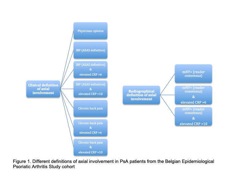
Methods: Patients originated from the Belgian Epidemiological Psoriatic Arthritis Study (BEPAS), a prospective multicentre cohort involving 17 Belgian rheumatology practices. Recruitment was from December 2012 until July 2014. Patients were included when fulfilling the Classification criteria for Psoriatic Arthritis (CASPAR). Axial involvement included several definitions (see figure 1). The treating did the clinical work up. Two calibrated central readers evaluated radiographic damage by assessing the modified Stoke Ankylosing Spondylitis Spinal Score (mSASSS) for the presence of syndesmophytes and assessed the modified New York criteria (mNY) for the presence of sacroiliitis. Readers assessed spinal and pelvic radiographs separately and were blinded for time sequence, clinical data, and information from other obtained images (radiographs of the hands and feet). Generalized estimating equation models on vertebral unit level were used, taking into account that multiple corners belong to a single patient. All definitions of axial involvement were evaluated, only odds ratios (OR) with a p< 0.05 were reported.
Results: In total 461 patients were included in BEPAS. Mean age was 52.79±12.29 years and 43.0% (n=198) were female; average disease duration was 8.5 ± 9.3 years and approximately 34% of the patients reported inflammatory axial pain. From 150 patients 2 years follow-up of spinal radiographs were obtained. From the clinical definitions of axial involvement none were associated with the development of syndesmophytes in 2 years. Axial involvement was associated with syndesmophyte formation when defined as ‘mNY+ reader consensus’ (OR 3.72 (CI 1.06-13.06)). Although few patients with PsA developed syndesmophytes after 2 years, conditional probability analysis suggests that those patients meeting the mNY criteria for axial involvement have a higher chance of developing syndesmophytes (Table 1).
Table 1. Conditional probability table for axial involvement defined as radiographic sacroiliitis (mNY+ reader consensus) and the development of syndesmophytes over 2 years follow-up.
| mNY+ reader consensus | New syndesmophytes at 2 years | n | P (newSYND/axial involvement) |
| 0 | 0 | 3446 | P (newSYND/0) = 34/3480 = 0.0098 |
| 0 | 1 | 34 | |
| 1 | 0 | 116 | P (newSYND/1) = 4/120 = 0.0333 |
| 1 | 1 | 4 | |
| 0=absent, 1=present |
Conclusion: The likelihood of syndesmophyte formation in PsA is low and is more likely to be associated with axial involvement determined radiographically, particularly in the context of high CRP.
To cite this abstract in AMA style:
de Hooge M, Ishchenko A, Steinfeld S, NZEUSSEU TOUKAP A, Elewaut D, Lories R, Van den bosch F, de Vlam K. Is Radiographic Axial Involvement Associated with Syndesmophyte Development After 2 Years in PsA Patients? [abstract]. Arthritis Rheumatol. 2022; 74 (suppl 9). https://acrabstracts.org/abstract/is-radiographic-axial-involvement-associated-with-syndesmophyte-development-after-2-years-in-psa-patients/. Accessed .« Back to ACR Convergence 2022
ACR Meeting Abstracts - https://acrabstracts.org/abstract/is-radiographic-axial-involvement-associated-with-syndesmophyte-development-after-2-years-in-psa-patients/

