Session Information
Date: Tuesday, November 9, 2021
Title: Osteoarthritis & Joint Biology – Basic Science Poster (1468–1479)
Session Type: Poster Session D
Session Time: 8:30AM-10:30AM
Background/Purpose: Subchondral bone marrow lesions (BMLs) are associated with symptoms and structural progression of knee OA. BMLs are characterized by diffuse hyperintensity on fat suppressed T2-weighted images. BMLs are commonly assessed using semi-quantitative (SQ) scoring systems by expert readers or volume quantification. Automated detection of BMLs using machine learning approaches may help in screening potential participants in clinical trials enriched for fast structural progression.
Methods: The dataset consists of 1,899 knee MRI exams from three sub-studies of the Osteoarthritis Initiative (OAI) with available baseline SQ readings: Foundation for NIH (FNIH) OA biomarker, Pivotal OAI MRI Analysis (POMA) incident radiographic knee OA, and POMA knee replacement. We trained a deep learning model (modified MRNet) using the sagittal intermediate-weighted (IW) fat-suppressed (FS) images. We dichotomized the MOAKS (MRI Osteoarthritis Knee Score) grades into presence or absence categories. The split was done by categorizing grades > 0 as presence and grades = 0 as absence. The whole data were randomly split into a training set (1524 exams) to train the model, a validation set (182 exams) to select the best model, and a test set (193 exams) to evaluate prediction performance. After the deep learning models were trained, we obtained probabilities of the existence of BMLs from IW images on each of 15 subregions in the femur and tibia. The logistic regression model was trained to combine the probabilities of different regions for IW images to output a probability of BMLs status for each subject overall. We utilized the area under the receiver operating characteristic curve (AUC) to assess the model’s performance and 95% confidence intervals of AUC to measure the variability of AUC. AUC values range from 0 to 1, with 1 indicating a 100% correct detection.
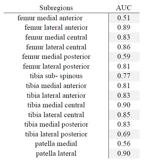
Results: The highest AUC value for IW images in the test set was 0.90 at two regions (tibia central medial and patella lateral). AUC values ranged from 0.51 to 0.89 in the test set for the other regions, see Table 1. At the subject level, the detection performance of the combined model achieved an AUC value of 0.87 with a 95% confidence interval of [0.82, 0.93].
Conclusion: We have developed and validated a fully automated deep learning framework to detect BMLs from MRI data. After the model had been trained, we applied the trained model to MRI data without MOAKS readings to predict the probability of BMLs status automatically. The prediction performance of detecting BMLs varied among different regions. Our method has some limitations, e.g., the prediction accuracy at the subject level is not as good as the accuracy for the individual subregions. In future studies, we will utilize images from other MRI sequences and additional MOAKS readings to improve our model’s performance. Automated classification of diffuse vs cystic BMLs and severity grading will be important in addition.
 Table 1. AUC values for the test set.
To cite this abstract in AMA style:
Liu S, Sun X, Roemer F, Ashbeck E, Bedrick E, Li Z, Guermazi A, Kwoh K. Automatic Detection of Bone Marrow Lesions from Knee MRI Data from the OAI Study [abstract]. Arthritis Rheumatol. 2021; 73 (suppl 9). https://acrabstracts.org/abstract/automatic-detection-of-bone-marrow-lesions-from-knee-mri-data-from-the-oai-study/. Accessed .« Back to ACR Convergence 2021
ACR Meeting Abstracts - https://acrabstracts.org/abstract/automatic-detection-of-bone-marrow-lesions-from-knee-mri-data-from-the-oai-study/
