Session Information
Session Type: Poster Session B
Session Time: 8:30AM-10:30AM
Background/Purpose: Bone marrow lesions (BML) are indicators for knee osteoarthritis (OA) and can be detected from magnetic resonance imaging (MRI). Due to the small size, low contrast, and various positions that BML may occur, it is challenging to automatically segment BMLs. Besides, not all patients with OA have BML, and for those patients with BML, not every slice has BML. The limited data becomes an obstacle to train machine learning models. In this work, we developed a novel strategy to synthesize MRI images with BML and trained deep learning models using the augmented dataset.
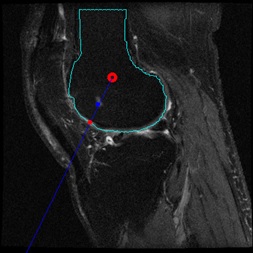
Methods: 300 cases from the Osteoarthritis Initiative (OAI) database were utilized in this study, where each case has sagittal intermediate weighted fat-suppressed (IWFS) sequence that is composed of a sequence of 36 MRI images. The BMLs in the dataset were manually delineated by domain experts as ground truth. We randomly separated the dataset into training, validation, and testing sets with 210/45/45 cases, respectively. To generate synthetic images with BML, we used images from the training set only. Three strategies were proposed. In strategy 1, for each image with BML in the training set, we selected a clean image without BML from the training set. We cropped out the BML from the original image and overlaid it on the clean image. To decide where to overlay it, we first calculated the center of the femur bone, the angle to the vertical direction, and the projected point on the bone edge in the original image, as shown in Figure 1. We also calculated the distance from the center to the lesion, and from the center to the bone edge. Then we applied the same distance percentage and same angle when overlaying the BML to the clean image. In strategy 2, everything is the same as strategy 1 except a fixed distance percentage was used to make sure the lesion is close to the bone boundary. In strategy 3, we introduced variations about the angle. A random angle from a range of -20 to +20 degrees from the vertical direction of the bone was applied when overlaying the BML. Figure 2 shows examples of the three strategies. After data generation, we first trained a deep learning model, called U-net, using the original BML images from the training set, as our baseline. Then for each strategy, a separate U-net model was trained, using the combination of original BML images plus the synthetic images.
Results: We reported the Dice coefficient and Similarity for each model in Table 1. An interesting finding is that for all the strategies, using 50% of the synthetic data produced higher accuracy than using 100% of the synthetic data. Therefore, we presented the results of using 50% of the synthetic data for each strategy. As Table 1 shows, the best accuracy was achieved by strategy 2, with more than 0.1 improvement in Dice and around 0.1 improvement in Similarity.
Conclusion: We developed an effective synthetic image generation method for BML segmentation. The accuracy was improved after adding the synthetic images to the training set. Future work could focus on refining the strategies for more realistic images and find the balanced combination between original images and synthetic images.
To cite this abstract in AMA style:
Michaely B, Zhang M, Shan J. Bone Marrow Lesion Segmentation Using Synthetic Data and Deep Learning Models [abstract]. Arthritis Rheumatol. 2021; 73 (suppl 9). https://acrabstracts.org/abstract/bone-marrow-lesion-segmentation-using-synthetic-data-and-deep-learning-models/. Accessed .« Back to ACR Convergence 2021
ACR Meeting Abstracts - https://acrabstracts.org/abstract/bone-marrow-lesion-segmentation-using-synthetic-data-and-deep-learning-models/


