Session Information
Session Type: Abstract Submissions (ACR)
Methods : Wb-MRIs of those 40 patients who reached the end of year 4 were scored for active inflammation (osteitis) and fatty lesions in the SI-joint quadrants and spine vertebral units (VUs). For this analysis we pooled the data of the patients who were continously treated with ETN for 3 consecutive years. Scoring was performed by two radiologists, blinded for treatment arm and MRI time point. Fatty lesions were scored for absence or presence according to our recently published score [2]. For this analysis, the presence or absence of osteitis and fatty lesions were only counted if both scorers agreed.
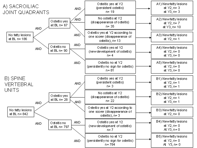
Results: New fatty lesions did not develop at all in sites (quadrants or VUs) where there was no osteitis at baseline.
New fatty lesions at year 2 (Y2) or Y3 primarily developed in SI-joint quadrants in which osteitis disappeared between baseline (BL) and Y2, this was the case in 28.6% quadrants (10 out of 35) (see Figure 1, section A2). In the spine only 1 VU developed a new fatty lesion in which osteitis disappeared between BL and Y2/Y3.
Persistent osteitis at Y2 or new development of osteitis (occurred rarely) was associated with a low rate of new development of fatty lesions, 16% (3/19) and 33% (1/3) for SI-joint and spine, respectively (A1, B1). New onset of osteitis at Y2 was not associated with new fatty lesions.
At baseline 74 SI-joint quadrants and 26 spine VUs showed fatty lesions. Until Y2 and Y3 only in 3 SI-joint quadrants and 0 VUs fatty lesions disappeared.
The mean spine fatty lesion scores were 1.1 (2.1) at BL, 1.4 (2.5) at Y2 and 1.3 (2.3) at Y3. The mean SI-joint fatty lesion scores were 4.6 (6.3), 5.2 (6.4) and 4.7 (6.3), respectively.
Conclusion : Our data indicate a strong relationship between presence of inflammation and new development of fatty lesions. Furthermore there was no increase of fatty lesions over continous treatment of axial SpA patients with etanercept over 3 years. . Whether this is predictive of stopping radiographic progression needs to be further investigated.
Figure 1: Development of new fatty lesions depending on the presence of inflammation on magnetic resonance imaging related to sacroiliac joint quadrants and spine vertebral units (VUs) with no fatty lesions at baseline (BL). Y= year; n= number of sites (SI-joint quadrants or spine vertebral units)
Referenences:
[1] Song I.-H. et al. Ann Rheum Dis. 2011 Apr;70(4):590-6.
[2] Song I.-H. et al. Ann Rheum Dis. 2011 Jul;70(7):1257-63.
Disclosure:
I. H. Song,
Pfizer, Merck Sharp Dohme/Schering Plough, AbbVie ,
5;
K. G. A. Hermann,
None;
H. Haibel,
AbbVie,MSD, Chugai,
8,
AbbVie, MSD,
5;
C. Althoff,
None;
D. Poddubnyy,
Merck Sharp Dohme/Schering Plough, AbbVie,
5;
J. Listing,
None;
A. Weiss,
None;
E. Lange,
Pfizer Inc,
3;
B. Freundlich,
Pfizer Inc,
9;
M. Rudwaleit,
Pfizer, Merck Sharp Dohme/Schering Plough, AbbVie, UCB,
5;
J. Sieper,
Pfizer, Merck Sharp Dohme/Schering Plough, AbbVie, UCB,
5.
« Back to 2013 ACR/ARHP Annual Meeting
ACR Meeting Abstracts - https://acrabstracts.org/abstract/relationship-between-active-inflammatory-lesions-in-the-spine-and-sacroiliac-joints-and-development-of-fatty-lesions-on-whole-body-mri-in-patients-with-early-axial-spondyloarthritis-long-term-d/

