ACR Meeting Abstracts

ACR Meeting Abstracts

  • Meetings
    • ACR Convergence 2024
    • ACR Convergence 2023
    • 2023 ACR/ARP PRSYM
    • ACR Convergence 2022
    • ACR Convergence 2021
    • ACR Convergence 2020
    • 2020 ACR/ARP PRSYM
    • 2019 ACR/ARP Annual Meeting
    • 2018-2009 Meetings
    • Download Abstracts
  • Keyword Index
  • Advanced Search
  • Your Favorites
    • Favorites
    • Login
    • View and print all favorites
    • Clear all your favorites
  • ACR Meetings

Abstract Number: 2750

Development and Preliminary Validation of a Novel Lung Ultrasound Interpretation Criteria for the Detection of Interstitial Lung Disease in Patients with Systemic Sclerosis

Robert Fairchild1, Diana Yang 2, Melody Chung 1, Laurel Sharpless 1, Shufeng Li 3 and Lorinda Chung 1, 1Stanford University, Palo Alto, CA, 2Santa Clara Valley Medical Center, Palo Alto, CA, 3Division of Immunology and Rheumatology, Department of Medicine, Stanford, Palo Alto, CA

Meeting: 2019 ACR/ARP Annual Meeting

Keywords: interstitial lung disease and ultrasound, Scleroderma, Systemic sclerosis

  • Tweet
  • Click to email a link to a friend (Opens in new window) Email
  • Click to print (Opens in new window) Print
Session Information

Date: Tuesday, November 12, 2019

Title: 5T089: Imaging of Rheumatic Diseases II (2750–2755)

Session Type: ACR Abstract Session

Session Time: 2:30PM-4:00PM

Background/Purpose: Interstitial lung disease (ILD) is a frequent complication of systemic sclerosis (SSc), and screening, characterization, and monitoring of disease activity are important for therapeutic decision-making and prognostication. High resolution computed tomography (HRCT) is the gold standard for ILD diagnosis. More recently, lung ultrasonography (LUS) has been proposed as a potential alternative imaging modality for ILD detection. In this study, we develop and test a novel LUS interpretation criteria for detecting SSc-ILD.

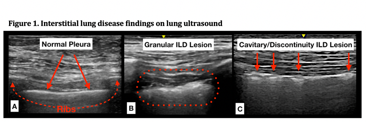
Methods: Patients with prevalent SSc and prior or planned HRCT within 3 months were prospectively enrolled and assessed by LUS. We included 14 lung positions and used a novel acquisition technique, collecting several seconds of real-time images rather than still images with the probe oriented in a sagittal rather than transverse orientation. This technique affords better visualization and tracking of pleural lesions with respiration. Prior studies identified B-line quantification, pleural irregularity, and pleural thickness as features sensitive for SSc-ILD. Although B-line quantification has been extensively studied, it is a less specific and artifactually based finding and highly dependent on ultrasound technique and machine settings. Given the high resolution of current US machines, granular appearing pleural thickening and pleural irregularity (defects in the pleural surface) were easy to visualize and highly reproducible (Figure 1). Using a combination of these features, we developed LUS interpretation criteria for SSc-ILD detection (Figure 2). These interpretation criteria were used to evaluate LUS examinations of our SSc cohort and were read by two independent readers (1 ultrasonographer and 1 non‑ultrasonographer) who were blinded to HRCT results and ILD diagnosis. The sensitivity and specificity for SSc-ILD detection was assessed and agreement was measured with Cohen’s Kappa statistic.

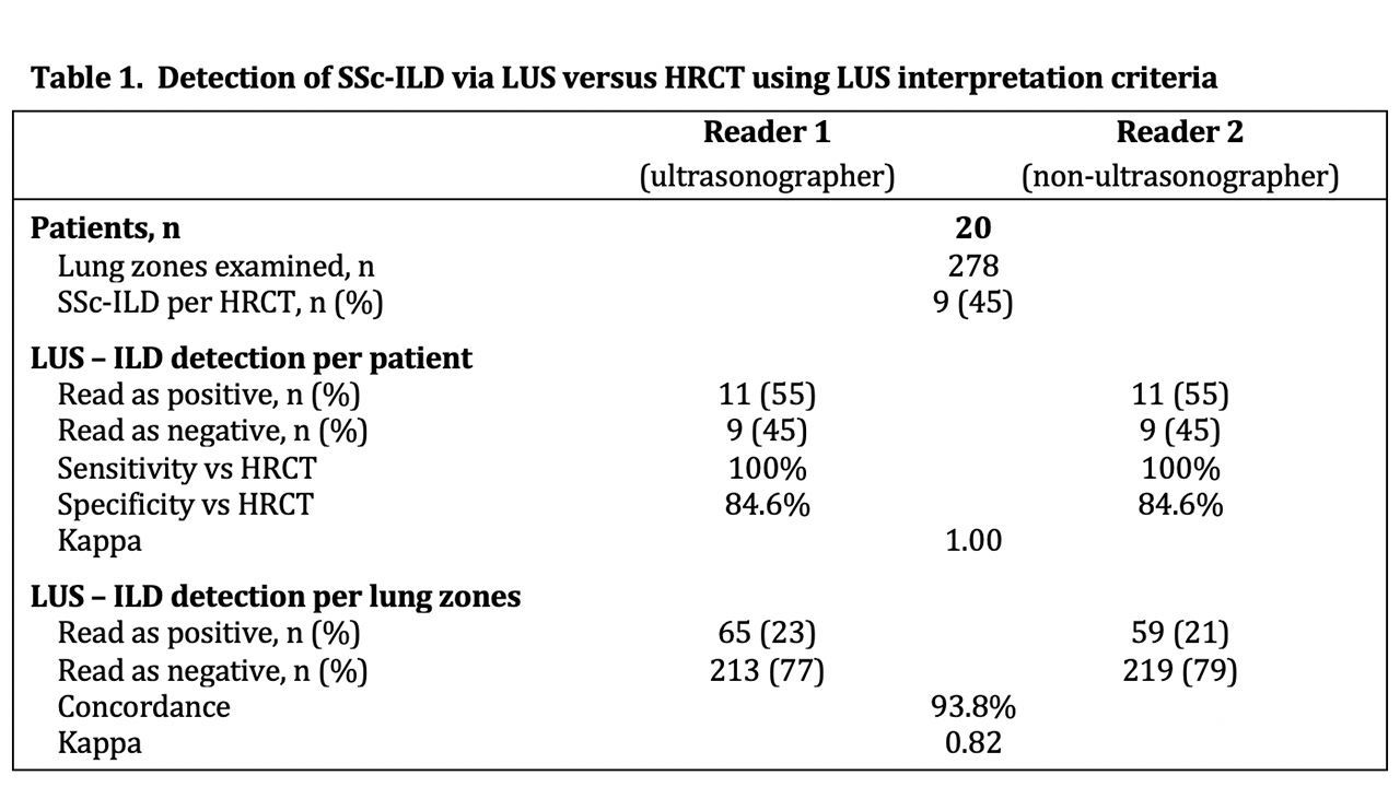
Results: We evaluated 20 SSc patients by LUS examination (278 lung zones) and HRCT. HRCT confirmed ILD in 9 patients (45%). LUS was read as positive for SSc-ILD in 11 patients (55%) with a sensitivity of 100% and specificity of 84.6% versus HRCT with perfect agreement between the two readers (κ= 1). Analysis by individual lung zones found excellent agreement between readers with 93.8% concordance and κ = 0.82 (Table 1).

Conclusion: We developed a novel ultrasound technique and interpretation criteria that are highly sensitive and specific for ILD detection in an SSc cohort. Our interpretation criteria are feasible and reliable, affording perfect agreement between ultrasonographer and non‑ultrasonographer readers for SSc-ILD detection. Moving forward we hope to validate these criteria in a larger cohort of patients with SSc, dermatomyositis and polymyositis.

-A- normal pleural surface with surrounding ribs, -B- granular pleural surface in ILD, -C- pleural discontinuities and cavitations in ILD. ILD=interstitial lung disease.

HRCT = high resolution computed tomography, LUS = lung ultrasound, ILD = interstitial lung disease, SSc-ILD = systemic sclerosis associated interstitial lung disease.


Disclosure: R. Fairchild, None; D. Yang, None; M. Chung, None; L. Sharpless, None; S. Li, None; L. Chung, BMS, 6, 9, Boehrenger-Ingelheim, 5, Boehringer Ingelheim, 5, Boehringer-Ingelheim, 5, Bristol-Myers Squib, 5, Eicos, 5, 6, 9, Eicos Steering Committee, 5, Mitsubishi Tanabe, 5, Reata, 5, 6, Reata DSMB, 5, Reatta, 5.

To cite this abstract in AMA style:

Fairchild R, Yang D, Chung M, Sharpless L, Li S, Chung L. Development and Preliminary Validation of a Novel Lung Ultrasound Interpretation Criteria for the Detection of Interstitial Lung Disease in Patients with Systemic Sclerosis [abstract]. Arthritis Rheumatol. 2019; 71 (suppl 10). https://acrabstracts.org/abstract/development-and-preliminary-validation-of-a-novel-lung-ultrasound-interpretation-criteria-for-the-detection-of-interstitial-lung-disease-in-patients-with-systemic-sclerosis/. Accessed .
  • Tweet
  • Click to email a link to a friend (Opens in new window) Email
  • Click to print (Opens in new window) Print

« Back to 2019 ACR/ARP Annual Meeting

ACR Meeting Abstracts - https://acrabstracts.org/abstract/development-and-preliminary-validation-of-a-novel-lung-ultrasound-interpretation-criteria-for-the-detection-of-interstitial-lung-disease-in-patients-with-systemic-sclerosis/

Advanced Search

Your Favorites

You can save and print a list of your favorite abstracts during your browser session by clicking the “Favorite” button at the bottom of any abstract. View your favorites »

All abstracts accepted to ACR Convergence are under media embargo once the ACR has notified presenters of their abstract’s acceptance. They may be presented at other meetings or published as manuscripts after this time but should not be discussed in non-scholarly venues or outlets. The following embargo policies are strictly enforced by the ACR.

Accepted abstracts are made available to the public online in advance of the meeting and are published in a special online supplement of our scientific journal, Arthritis & Rheumatology. Information contained in those abstracts may not be released until the abstracts appear online. In an exception to the media embargo, academic institutions, private organizations, and companies with products whose value may be influenced by information contained in an abstract may issue a press release to coincide with the availability of an ACR abstract on the ACR website. However, the ACR continues to require that information that goes beyond that contained in the abstract (e.g., discussion of the abstract done as part of editorial news coverage) is under media embargo until 10:00 AM ET on November 14, 2024. Journalists with access to embargoed information cannot release articles or editorial news coverage before this time. Editorial news coverage is considered original articles/videos developed by employed journalists to report facts, commentary, and subject matter expert quotes in a narrative form using a variety of sources (e.g., research, announcements, press releases, events, etc.).

Violation of this policy may result in the abstract being withdrawn from the meeting and other measures deemed appropriate. Authors are responsible for notifying colleagues, institutions, communications firms, and all other stakeholders related to the development or promotion of the abstract about this policy. If you have questions about the ACR abstract embargo policy, please contact ACR abstracts staff at [email protected].

Wiley

  • Online Journal
  • Privacy Policy
  • Permissions Policies
  • Cookie Preferences

© Copyright 2025 American College of Rheumatology