Session Information
Session Type: Poster Session (Monday)
Session Time: 9:00AM-11:00AM
Background/Purpose: Background/Purpose: Discriminating inflammatory pain from non-inflammatory pain is critical to determine therapeutic strategy in inflammatory arthritis (IA) such as rheumatoid arthritis (RA) and spondyloarthritis (SpA). Previous studies showed that resting-state functional MRI (rs-fMRI) could detect abnormal functional connectivity (FC) of brain areas related to chronic pain and could predict disease course as in chronic lumbar pain. However, the difference of FC between inflammatory and non-inflammatory pain is still unknown. This study aimed to clarify the abnormal FC related to inflammatory pain and whether it could predict the response of treatment in IA.
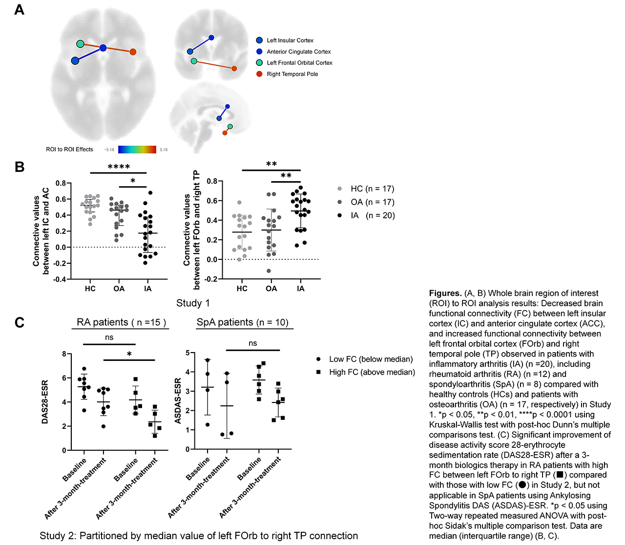
Methods: Methods: IA patients requiring biologics (n = 46; RA 28, SpA 18) and healthy controls (HCs, n = 17) underwent rs-fMRI in Hokkaido University Hospital. For controls of non-inflammatory pain, rs-fMRI dataset of patients with osteoarthritis (OA) (n = 17) was obtained from OpenNEURO (doi: 10.18112/openneuro.ds000208.v1.0.0). IA patients were split into two groups (Study 1 or Study 2), depending on whether or not the patients had clinical follow-up data after biologics therapy. In Study 1, to identify the abnormal FC related to inflammatory pain, rs-fMRI-derived FC values to whole brain connectivity were retrospectively analyzed in IA patients (n = 20; RA 12, SpA 8) by comparing with OA patients and HCs (n = 17, respectively). In Study 2, IA patients treated with biologics were prospectively evaluated with disease activity score 28-erythrocyte sedimentation rate (DAS28-ESR) and Ankylosing Spondylitis DAS-ESR (ASDAS-ESR) in RA patients (n = 15) and SpA patients (n = 10), respectively before treatment and after a-3-month therapy. We then analyzed the association between the detected FC and disease activity.
Results: Results: From the whole brain analyses in Study 1, significant connective values between left insula cortex (IC) and anterior cingulate cortex (ACC), and left frontal orbital cortex (FOrb) and right temporal pole (TP) were found in IA patients compared with OA patients and HCs (Figure. 1A, B). In Study 2, the connectivity values of left FOrb and right TP partitioned by median value of IA patients could discriminate significant improvement of DAS28-ESR after treatment in RA patients, although there was no association with the improvement of ASDAS-ESR in SpA patients (Figure. 1C).
Conclusion: Conclusions: RA and SpA patients shared neurobiologic features of the abnormal functional connections between left IC and ACC, and left FOrb and right TP, which are brain regions involving pain processing and decision making. High FC value between left FOrb and right TP would predict the superior therapeutic outcomes regarding disease activity in patients with RA, suggesting that this abnormal FC would be associated with inflammatory pain.
To cite this abstract in AMA style:
Abe N, Fujieda Y, Asou K, Karino K, Kono M, Narita H, Kato M, Tha K, Oku K, Amengual O, Yasuda S, Atsumi T. Resting-State Functional Connectivity of Pain Processing Brain Region Associated with Therapeutic Response to Biologics in Rheumatoid Arthritis and Spondyloarthritis [abstract]. Arthritis Rheumatol. 2019; 71 (suppl 10). https://acrabstracts.org/abstract/resting-state-functional-connectivity-of-pain-processing-brain-region-associated-with-therapeutic-response-to-biologics-in-rheumatoid-arthritis-and-spondyloarthritis/. Accessed .« Back to 2019 ACR/ARP Annual Meeting
ACR Meeting Abstracts - https://acrabstracts.org/abstract/resting-state-functional-connectivity-of-pain-processing-brain-region-associated-with-therapeutic-response-to-biologics-in-rheumatoid-arthritis-and-spondyloarthritis/

