Session Information
Session Type: ACR Poster Session B
Session Time: 9:00AM-11:00AM
Background/Purpose: Osteoarthritis (OA) is a rheumatic disease characterized by articular cartilage degradation. On its early stages, OA is asymptomatic and its current gold-standard diagnosis (X-Rays), focused on changes on the adjacent tissue – bone – presents a limitation to the early diagnosis of this disease. Raman spectroscopy (RS) has been recently described as a non-invasive tool to detect molecular changes in biological tissues, producing a unique fingerprint. For this reason, its application for the diagnosis of different diseases offers high potential. Beyond its clinical application, RS offers value as a molecular quantification technique for tissue engineered cartilage characterization. The aim of this work was to evaluate the potential of RS for the early diagnosis of OA.
Methods: Human hip cartilage explants (n=14), from healthy (H) and OA donors, with Kellgren-Lawrence (K-L) radiological grades from 0 to IV, were obtained after informed consent. Raman analysis was performed on fresh tissue, using a Bruker RFS100 Spectrometer with a Nd:YaG laser (ʎ=1064). Main peaks were assigned according to literature3, following their area’s measurement after a normalization process. One-way ANOVA statistical analysis was performed and differences considered significant for p<0.05. We further analyzed correlations (Pearson’s coefficient) between peaks and K-L grade.
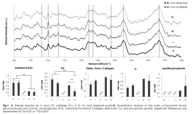
Results: RS cartilage spectra (Fig.1A) revealed the following assignments: 1245-1270cm-1 amide III doublet (random coil and α-helix collagen), 1063cm-1 (sulfated glycosaminoglycans, GAGs), 1377cm-1 (proteoglycans, PG), 1450cm-1 (nonspecific signal of lipids and proteins) and 1668cm-1 (carbonyl group in proteins). For higher K-L grades, a peak appeared at 960cm-1 (apatite phosphate), related to tissue mineralization. After quantitative analysis (Fig.1B) we observed the main molecular changes: GAGs and PG peaks showed a significant decrease with OA severity (p<0.01), supported by high correlation coefficients (R2=0.7361 and R2=0.7999, respectively), related to GAGs’ degradation; an increase in 1245/1270 ratio (defective/functional collagen) could reveal collagen arrangement loss, although there was a low correlation vs K-L (R2=0.3764); an indirect lipid index (IL), calculated as A1450/A1668, showed an increase of lipids in OA tissues.
Conclusion: RS analysis revealed a hip cartilage molecular fingerprint. Variations found between H and OA tissue are representative of the molecular changes during OA progression. A set of parameters is suggested as an optical biomarker panel: defective/functional collagen (1245-1270cm-1), GAGs (1063cm-1), PG (1377cm-1) and IL (1450/1668).
To cite this abstract in AMA style:
Casal Beiroa P, Burguera EF, Hermida-Gómez T, Goyanes N, Oreiro N, González P, Blanco FJ, Magalhaes J. Optical Biomarkers for the Early Diagnosis of Osteoarthritis [abstract]. Arthritis Rheumatol. 2018; 70 (suppl 9). https://acrabstracts.org/abstract/optical-biomarkers-for-the-early-diagnosis-of-osteoarthritis/. Accessed .« Back to 2018 ACR/ARHP Annual Meeting
ACR Meeting Abstracts - https://acrabstracts.org/abstract/optical-biomarkers-for-the-early-diagnosis-of-osteoarthritis/

