Session Information
Session Type: ACR Poster Session C
Session Time: 9:00AM-11:00AM
Background/Purpose: Serum uric acid (sUA) is a useful indicator of the risk of developing gout. However, most patients with elevated sUA levels do not have gout. Dual-energy computed tomography (DECT) scan is a relatively sensitive and specific imaging tool used to measure and visualize urate deposits in the joints. Prior research shows presence of subclinical urate deposits in nearly one-quarter of patients with severe hyperuricemia (i.e., sUA >9 mg/dl) without gout. We conducted a cross-sectional study to identify urate deposits using DECT and examine whether an association exists between sUA levels and total volume of urate deposits in patients with asymptomatic hyperuricemia.
Methods: We recruited adults aged ≥40 years with sUA levels ≥6.5 mg/dl and metabolic syndrome according to the National Cholesterol Education Program–Adult Treatment Panel III criteria. We excluded patients with gout, end-stage renal disease, renal replacement therapy, active malignancy, and use of xanthine oxidase inhibitors, colchicine or probenecid. All patients underwent a measurement of sUA level and DECT scan of a foot. A fellowship trained MSK radiologist processed and reviewed the DECT images and determined total volume of urate deposits. We used logistic regression to assess the association between sUA and presence of urate deposits and linear regression to examine the association between sUA and total volume of urate deposits.
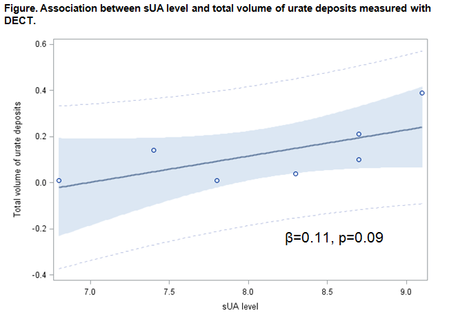
Results: A total of 46 subjects participated in this study. The mean age was 62 (±8) years, 41% were male and mean sUA level was 7.8 (±1.0) mg/dl. Seven of 46 (15%) patients had urate deposits in the feet. The mean total volume of urate deposits was 0.13 (±0.14) cm3. On univariable analysis, age had significant association with presence of urate deposits but sUA, male sex, body mass index, presence of diabetes, and renal function did not (Table). sUA had a modest linear association (β coefficient=0.11, p=0.09), albeit statistically not significant, with total volume of urate deposits (Figure).
Conclusion: In this cross-sectional study, 15% of patients with elevated sUA but no known gout had urate deposits in the foot DECT scan. There were no clear patient factors other than older age associated with presence of urate deposits on DECT scans. While the clinical significance of these urate deposits is unclear, it is possible that these patients may develop gouty arthritis or they may have “resistance” to reacting against urate depositis. Further research on why certain patients with hyperuricemia develop gout may present important clues to gout prevention.
To cite this abstract in AMA style:
Wang P, Smith S, Garg R, Lu F, Wohlfahrt A, Campos A, Vanni K, Yu Z, Solomon DH, Kim SC. Identification of Urate Deposits in Patients with Asymptomatic Hyperuricemia Using a Dual-Energy CT Scan [abstract]. Arthritis Rheumatol. 2017; 69 (suppl 10). https://acrabstracts.org/abstract/identification-of-urate-deposits-in-patients-with-asymptomatic-hyperuricemia-using-a-dual-energy-ct-scan/. Accessed .« Back to 2017 ACR/ARHP Annual Meeting
ACR Meeting Abstracts - https://acrabstracts.org/abstract/identification-of-urate-deposits-in-patients-with-asymptomatic-hyperuricemia-using-a-dual-energy-ct-scan/


