ACR Meeting Abstracts

ACR Meeting Abstracts

  • Meetings
    • ACR Convergence 2024
    • ACR Convergence 2023
    • 2023 ACR/ARP PRSYM
    • ACR Convergence 2022
    • ACR Convergence 2021
    • ACR Convergence 2020
    • 2020 ACR/ARP PRSYM
    • 2019 ACR/ARP Annual Meeting
    • 2018-2009 Meetings
    • Download Abstracts
  • Keyword Index
  • Advanced Search
  • Your Favorites
    • Favorites
    • Login
    • View and print all favorites
    • Clear all your favorites
  • ACR Meetings

Abstract Number: 1452

  18 F-FDG PET Imaging: An In Vivo quantitative Drug Screening Tool for Novel Antiinflammatory Therapies

Siba P. Raychaudhuri1, Smriti K. Raychaudhuri2, Anupam Mitra3 and Abhijit Chaudhari4, 1Davis, CA, 2Rheumatology/Immunology, VA Sacramento Medical Center, Davis, CA, 3Dermatology, UC Davis School of Medicine, Davis, CA, 4Radiology, UC Davis School of Medicine, Sacramento, CA

Meeting: 2016 ACR/ARHP Annual Meeting

Date of first publication: September 28, 2016

Keywords: Animal models, Arthritis, drug treatment and imaging techniques

  • Tweet
  • Click to email a link to a friend (Opens in new window) Email
  • Click to print (Opens in new window) Print
Session Information

Date: Monday, November 14, 2016

Title: Rheumatoid Arthritis – Animal Models - Poster II

Session Type: ACR Poster Session B

Session Time: 9:00AM-11:00AM

Background/Purpose: Collagen induced arthritis (CIA) mouse model is used for screening of new drugs for autoimmune arthritis. The conventional read outs of this model are clinical and histological scores. These read-outs have many limitations including (i) longitudinal studies in the same mouse cannot be performed (ii) clinical and histological scores are subjected to observer bias (iii) in vivo cellular events cannot be captured in its native environment. To meet these unmet needs here we have evaluated the utility of 18F-FDG PET imaging in the CIA model.     

Methods:  Briefly, arthritis was induced using bovine type II collagen in 8-week old male DBA/1J mice (n=30), out of which 20 mice developed arthritis. The disease was allowed to progress till day 45 (pre-treatment time point- Fig 1), 5 mice were sacrificed for histological studies; 10 mice were treated with  i.p anti-mouse TNF-α antibody (CNTO5048, Janssen Biotech, USA) every alternate day (300 ug/dose) for next 10 days, and 5 mice received sterile PBS (vehicle control). Mice were scored clinically and had 18F-FDG PET scan on day 0, 28, 45 and 56.  The maximum 18F-FDG uptake (standardized uptake value normalized by body weight) was determined for each major joints to generate a comprehensive PET score (PS).  

Results: All data represented as median with range.  The median clinical score (CS) of the most affected limb in the vehicle treated and untreated gradually increased to 2 (range:1-2) at day 28 (p<0.01), 2 (range:2-3) at day 45 (p<0.01) and 3 (range:2-3) at day 56 (p<0.01) compared to day 0. The clinical observation was confirmed by histological scores (HS). Compared to day 0 (HS=0), the median HS on day 28 was 2 (range:1-3), day 45 was 4 (range:2-4) and day 56 was 4 (range:4-5). The PS on day 0 was 1.02 (range:0.85-1.05). PS increased to median values of 1.52 (range:1.14-2.01) on day 28 (p<0.05), 3.51 (range:1.23-4.76) on day 45 (p<0.05) and 1.76 (range:1.45-2.78) on day 56 (p<0.05). CNTO5048 treatment significantly reduced the median CS to 1 (range:0-2, p<0.01) and HS to 1 (range:1-2, p<0.01) compared to their respective pre-treatment levels (CS:2, HS:4). Similar to CS and HS, PS on day 56 was significantly low (median=0.97, range=0.62-2.12, p<0.01) compared to pre-treatment level (day 45) (median=3.51, range=1.23-4.76) (Fig 1).

Conclusion: Here we have validated in the CIA mouse model that 18F-FDG PET is an in vivo preclinical drug screening tool for antiinflammatory therapies. The use of this imaging modality will allow longitudinal quantitative evaluation response in the same mouse.    

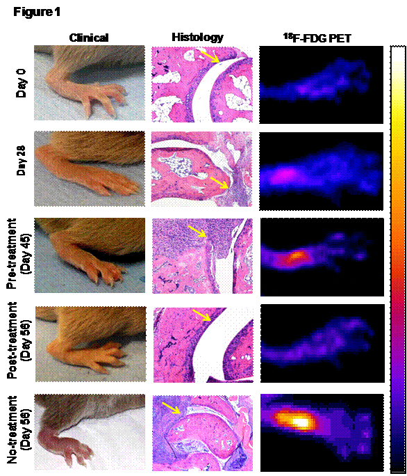
Figure 1: 18F-FDG PET imaging, a quantative in vivo pre-clinical drug screening tool. 18F-FDG PET imaging effectively corelated with the both clinical and histopayhological (yellow arrows) therapuetic response of the anti-TNF-α (CNTO5048) antibody. The pseudo-color in PET indicates higher cellular metabolic activity, which resiprocates with the degree of inflammation.   


Disclosure: S. P. Raychaudhuri, None; S. K. Raychaudhuri, None; A. Mitra, None; A. Chaudhari, None.

To cite this abstract in AMA style:

Raychaudhuri SP, Raychaudhuri SK, Mitra A, Chaudhari A.   18 F-FDG PET Imaging: An In Vivo quantitative Drug Screening Tool for Novel Antiinflammatory Therapies [abstract]. Arthritis Rheumatol. 2016; 68 (suppl 10). https://acrabstracts.org/abstract/18-f-fdg-pet-imaging-an-in-vivo-quantitative-drug-screening-tool-for-novel-antiinflammatory-therapies/. Accessed .
  • Tweet
  • Click to email a link to a friend (Opens in new window) Email
  • Click to print (Opens in new window) Print

« Back to 2016 ACR/ARHP Annual Meeting

ACR Meeting Abstracts - https://acrabstracts.org/abstract/18-f-fdg-pet-imaging-an-in-vivo-quantitative-drug-screening-tool-for-novel-antiinflammatory-therapies/

Advanced Search

Your Favorites

You can save and print a list of your favorite abstracts during your browser session by clicking the “Favorite” button at the bottom of any abstract. View your favorites »

All abstracts accepted to ACR Convergence are under media embargo once the ACR has notified presenters of their abstract’s acceptance. They may be presented at other meetings or published as manuscripts after this time but should not be discussed in non-scholarly venues or outlets. The following embargo policies are strictly enforced by the ACR.

Accepted abstracts are made available to the public online in advance of the meeting and are published in a special online supplement of our scientific journal, Arthritis & Rheumatology. Information contained in those abstracts may not be released until the abstracts appear online. In an exception to the media embargo, academic institutions, private organizations, and companies with products whose value may be influenced by information contained in an abstract may issue a press release to coincide with the availability of an ACR abstract on the ACR website. However, the ACR continues to require that information that goes beyond that contained in the abstract (e.g., discussion of the abstract done as part of editorial news coverage) is under media embargo until 10:00 AM ET on November 14, 2024. Journalists with access to embargoed information cannot release articles or editorial news coverage before this time. Editorial news coverage is considered original articles/videos developed by employed journalists to report facts, commentary, and subject matter expert quotes in a narrative form using a variety of sources (e.g., research, announcements, press releases, events, etc.).

Violation of this policy may result in the abstract being withdrawn from the meeting and other measures deemed appropriate. Authors are responsible for notifying colleagues, institutions, communications firms, and all other stakeholders related to the development or promotion of the abstract about this policy. If you have questions about the ACR abstract embargo policy, please contact ACR abstracts staff at [email protected].

Wiley

  • Online Journal
  • Privacy Policy
  • Permissions Policies
  • Cookie Preferences

© Copyright 2025 American College of Rheumatology