Session Information
Session Type: ACR Poster Session A
Session Time: 9:00AM-11:00AM
Background/Purpose: Traditionally, regions of the knee that are assessed in clinical trials are selected based on anatomy or responsiveness to change. However, it is unclear whether there are regions of the articular cartilage that are changing together over time. The purpose of the study was to visualize and explore which regions of cartilage change together longitudinally.
Methods: To determine the locations of greatest longitudinal cartilage change, we measured a convenience sample of 100 knees with baseline and 24-month MRIs from the Osteoarthritis Initiative. Sample included knees that were primarily Kellgren-Lawrence grades 2 or 3, with few progressing over 24 months. One reader (MZ) (intraclass correlation coefficient [ICC] 3,1 model > 0.86 at baseline) used customized software to measure the Cartilage Damage Index (CDI) in the medial and lateral compartments of the femur, tibia, and patellar cartilage on paired baseline and 24-month double echo steady state MRIs. For the whole knee CDI, cartilage thickness is measured in 12 informative locations for the medial and lateral patella, and in 9 locations for the medial and lateral femur and tibia (total of 60 CDI sites per knee). Using changes in CDI measures at each location over 24 months, we performed a factor analysis to identify sites that changed together. Factors with an eigenvalue > 1 were retained.
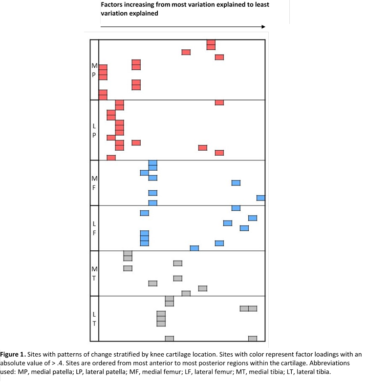
Results: The factor analysis produced 20 factors (Figure 1) accounting for 74% of the variance in CDI changes. To visualize the findings of the factor analysis, we created a figure that identifies the CDI points with factor loadings greater than or equal to 0.4 based on their anatomic location within the knee (medial and lateral patella, femur, and tibia). The factors are listed from greatest to smallest eigenvalues, or those explaining the largest percent of variability to those explaining the smallest percent of variability. Notably, the first 3 factors, that account for 23% of the total variation, only involved CDI locations in the patella. Surprisingly, very few of the factors (4) represent a combination of the articular surfaces that anatomically are adjacent to one another. Many of the factors (13) only involve one articular surface.
Conclusion: Factor analysis on this sample suggests that much of the variation in CDI change measures comes from regions on the patella. The patellofemoral joint is often overlooked in OA research, but our results suggest more research on this region of the knee is needed. Also, we found that it is uncommon for articular cartilage to change in conjunction with the adjacent articular surface (e.g. medial tibia cartilage does not necessarily change when the medial femur cartilage changes). These findings suggest that new strategies that evaluate change in cartilage may improve our understanding of OA progression.
To cite this abstract in AMA style:
Canavatchel AR, Lo GH, LaValley MP, Zhang M, Driban JB, Price LL, Miller E, Eaton C, McAlindon TE. Visualizing Different Patterns of Cartilage Change: A Two-Year Study of Data from the Osteoarthritis Initiative [abstract]. Arthritis Rheumatol. 2016; 68 (suppl 10). https://acrabstracts.org/abstract/visualizing-different-patterns-of-cartilage-change-a-two-year-study-of-data-from-the-osteoarthritis-initiative/. Accessed .« Back to 2016 ACR/ARHP Annual Meeting
ACR Meeting Abstracts - https://acrabstracts.org/abstract/visualizing-different-patterns-of-cartilage-change-a-two-year-study-of-data-from-the-osteoarthritis-initiative/

