Session Information
Session Type: ACR Poster Session A
Session Time: 9:00AM-11:00AM
Background/Purpose: Loss of joint space width (JSW) on x-ray is the recommended standard to define osteoarthritis progression. However, both cartilage and meniscal damage contribute to JSW loss, yet neither are seen on radiographs. The extent to which JSW loss on x-ray reflects cartilage damage vs. meniscal damage/extrusion is unknown. Our objective was to evaluate the impact of medial cartilage and meniscal damage/extrusion 1 year prior to incident radiographic osteoarthritis (ROA) on the subsequent trajectory of medial JSW.
Methods: Knees (n=327) from the OAI that developed incident ROA (IROA) through 48 months of follow-up were identified based on Kellgren-Lawrence grade (KLG) ≥2. MRI Osteoarthritis Knee Score (MOAKS) was used to assess cartilage damage (≥1.1 vs. <1.1) and meniscal damage/extrusion (i.e., tears and maceration [≥2 vs. <2] or extrusion [≥3mm vs. <3mm]) in the year prior to incident ROA. Annual fixed JSW (fJSW) (x=0.250 mm) on x-ray was assessed over three years of follow-up. The sample included n=1,289 fJSW measures in 327 knees from 301 participants between 2 years prior to IROA and up to 2 years after IROA detection. Trajectories of mean medial fJSW and change in medial fJSW, with 95% confidence intervals (CI), were estimated using mixed models with participant and knee treated as random effects.
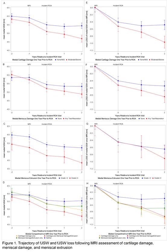
Results: As seen in Figure 1, knees with medial cartilage damage one year prior to IROA (compared to knees without damage) had a significantly lower mean medial fJSW in the three years following, with a difference in mean fJSW of 0.393mm [95%CI: 0.182, 0.604; p<0.0003] one year later, 0.454mm [95%CI: 0.233, 0.676; p<0.0001] two years later, and 0.754mm [95%CI: 0.508, 1.000; p<0.0001] three years later. Similarly, knees with medial meniscal damage one year prior to IROA (compared to those without damage) had significantly lower mean medial fJSW in the 3 years following, with a difference in mean fJSW of 0.238 [95%CI: 0.002, 0.474; p<0.0483] one year later, 0.288 [95%CI: 0.041, 0.534; p<0.0223] 2 years later, and 0.448 [95%CI: 0.180, 0.716; p<0.0011] 3 years later. Although those with meniscal extrusion had less fJSW one year prior to IROA, there was no significant difference in trajectory of fJSW loss for knees with meniscal extrusion vs. those without. There was no significant difference in the trajectory of fJSW loss among those with cartilage and meniscal damage/ extrusion vs. those with only cartilage damage.
Conclusion: Knees with medial tibiofemoral cartilage damage in the year prior to IROA, with or without meniscal damage/extrusion, had significant loss of mean medial fJSW over the following 3 years. Meniscal extrusion was associated with differences in concurrently measured fJSW, but not the future trajectory of fJSW. While loss of fJSW reflects both cartilage and meniscal damage, the magnitude of the differences suggests cartilage damage is the predominant factor in the future loss of fJSW.
To cite this abstract in AMA style:
Kwoh CK, Roemer F, Ashbeck EL, Ratzlaff C, Duryea J, Guermazi A. MRI-Detected Cartilage Damage, Meniscal Damage, and Meniscal Extrusion Prior to Incident Radiographic Osteoarthritis and the Subsequent Trajectory of Joint Space Loss [abstract]. Arthritis Rheumatol. 2016; 68 (suppl 10). https://acrabstracts.org/abstract/mri-detected-cartilage-damage-meniscal-damage-and-meniscal-extrusion-prior-to-incident-radiographic-osteoarthritis-and-the-subsequent-trajectory-of-joint-space-loss/. Accessed .« Back to 2016 ACR/ARHP Annual Meeting
ACR Meeting Abstracts - https://acrabstracts.org/abstract/mri-detected-cartilage-damage-meniscal-damage-and-meniscal-extrusion-prior-to-incident-radiographic-osteoarthritis-and-the-subsequent-trajectory-of-joint-space-loss/

